A B
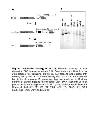
C24 D mbl C24 M C24 D mbl::aac M145 D mbl-2 D mbl1 M. A B. B ........ 5014 bp ......... B. M145 D mbl::aac D mbl. 2452. sco2450. mbl. red. l. B ...........5657 bp ............... B. 2452. sco2450. aac. frt. B ....4325 bp ..... B. 2452.

A B
E N D
Presentation Transcript
C24Dmbl C24 M C24Dmbl::aac M145 Dmbl-2 Dmbl1 M A B B ........ 5014 bp ......... B M145 Dmbl::aac Dmbl 2452 sco2450 mbl red l B ...........5657 bp ............... B 2452 sco2450 aac frt B ....4325 bp ..... B 2452 sco2450 Fig. S1.Inactivation strategy of mbl. A, Schematic drawing. mbl was deleted by PCR targeting of cosmid C24 (Redenbach et al., 1996) in a two step process, first replacing mbl by an aac cassette and subsequently deleting aac by FRT recombination, leaving a 81 bp scar sequence (hatched bar) in the chromosome. B,Mutant genotype was confirmed by Southern blotting of BamHI digested chromosomal DNA. DNA fragments used as probes are drawn as a grey line in A. M, Dig VII Molecular weight Standard, Roche (81, 359, 492, 710, 718, 992, 1164, 1482, 1515, 1882, 1953, 2799, 3639, 4899, 6106, 7427, and 8576 bp)
A B C M145 Dsco6166 M E Bg H B ............... 3421 bp ................. B B 6167 6166 sco6167 M145 Dsco6166 recA aphII B ......... Bg .. 2608 bp ... B B pKO6166 sco6167 Fig. S2. Inactivation of sco6166. A) Schematic drawing of the knock out plasmid, carrying upstream and downstream regions. Relevant restriction sites are indicated. E: EcoRI, Bg: BglII, H: HindIII. B) Schematic maps of the wildtype and the Dsco6166 mutant. A grey bar indicates the probe used in Southern blotting (C). The sizes of the hybridysing fragments are given. Relevant BamHI sites (B) are indicated. C) Southern blot of BamHI digested total DNA of M145 and the Dsco6166 mutant. M, DigVII Standard, Roche.
105 104 DmreB Dmbl DmreB/mbl M145 Dsco6166 Fig. S3.Sporulation defect of S. coelicolor mutants defective in mreB homologues on MS agar + 10.3 % sucrose. Approximately 105 or 104spores of M145, DmreB, Dmbl, DmreB/mbl and Dsco6166 were spotted on MS agar containing 10.3 % sucrose and incubated for 5 days at 30°C before taking the photograph. Whereas the wildtype M145 and the Dsco6166 mutant were able to sporulate, DmreB, Dmbl, and DmreB/mbl were affected in sporulation.
M145 Dmbl Fig. S4.Transmission electron micrographs of M145 and the Dmbl mutant. Thin sections of spores of S. coelicolor M145 (A) and its mbl gene replacement mutant (B) were analysed at high resolution by transmission electron microscopy. Integrity of the Dmbl spore walls is compromised, resulting in aberrant spores with a diffuse wall. Since the Dmbl spores are more sensitive than the wildtype spores, we can not exclude that the appearance of the spore surface was affected by the fixation/staining process. Bar = 500 nm.
phase contrast SCO6166-mCherry Fig. S5. Disperse fluorescence of SCO6166-mCherry in the substrate mycelium of S. coelicolor M145. A non-replicative plasmid carrying the sco6166-mcherry fusion gene was integrated into the chomosomal sco6166 gene of M145 via homologous recombination. Culture was grown on LB agar for two days. Bar, 4 µm.
phase contrast MreB-eGFP Dmbl Dpbp2 Fig. S6. Localization of MreB-eGFP to the spore periphery in mutants Dmbl and Dpbp2. Cultures were grown on MS agar for three days. Bar, 4 µm. MreB-eGFP localization underneath the spore membrane is not affected by the absence of Mbl or PBP2. Fig. S3
M145 48 h M145::pAH5 48 h M145::pAH5 72h DmreB::pAH5 48h DmreB::pAH5 72h H2O hrdB mbl Fig. S7. Transcriptional analysis of the mbl-mcherry fusion gene in the DmreB deletion mutant. Cultures were grown on cellophane discs on MS agar at 30°C and harvested after 48 h or 72 h. The amounts of transcripts were compared by PCR. Expression of mbl-mcherry was detectable in the DmreB mutant in considerable amounts. Transcription of the housekeeping hrdB gene and transcription of mbl in M145 (48 h) were used as controls.