MOLECULAR DIAGNOSTICS
MOLECULAR DIAGNOSTICS. Introduction: Definitions, Problematics, Examples Immunological Diagnostic Methods DNA Diagnostics Methods Bacterial Biosensors. Molecular Diagnostics. The success of modern medicine depends on the detection of specific molecules e.g. Viruses Bacteria Fungi

MOLECULAR DIAGNOSTICS
E N D
Presentation Transcript
MOLECULAR DIAGNOSTICS Introduction: Definitions, Problematics, Examples Immunological Diagnostic Methods DNA Diagnostics Methods Bacterial Biosensors
Molecular Diagnostics • The success of modern medicine depends on the detection of specific molecules e.g. • Viruses • Bacteria • Fungi • Parasites • Proteins • In water, plants, soil and humans.
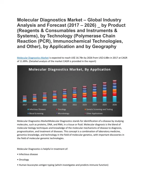
Molecular Diagnostics are Transforming Medicine Molecular diagnostics is >$3 billion market WW and growing at >20% annually Recurrence monitoring Drug selection Disease detection Disease predisposition Key questions Pre-natal testing “Is the baby healthy? “ “What diseases is this patient at risk for?” “Has this patient a disease?” “What drugs should I prescribe?” “How has the disease returned?” -> Need for Molecular tests
Molecular Diagnostics Characteristics of a Detection System • A good detection system should have 3 qualities: • Sensitivity • Specificity • Simplicity • Sensitivity means that the test must be able to detect very small amounts of target even in the presence of other molecules. • Specificity: the test yields a positive result for the target molecule only. • Simplicity: the test must be able to run efficiently and inexpensively on a routine basis.
MassARRAY Diagnostics Are Being Developed For Multiple Disease Areas Genetic Testing • High throughput testing for genetic disorders including single nucleotide polymorphisms (SNPs) markers, insertions, deletions • Examples: Factor II, Factor V, CFTR Prenatal Diagnostics • Non-invasive detection of fetal diseases • Examples: Down syndrome, cystic fibrosis • Progress is being made in all of these areas • Each of these areas are commercially attractive • In some cases, the MassARRAY platform is uniquely qualified for specific tests • More tests will be added to the platform as these tests are rolled out Oncology • Early diagnosis of cancer • Example: circulating tumor DNA TransplantationMedicine • Non-invasive, early detection of organ rejection • Example: urine testing for kidney rejection • Pathogen identification and early detection • Examples: identification of multi drug resistant mycobacteria, early detection of drug-resistant viral strains, e.g. HIV, HBV, HCV Infectious Disease
Molecular Diagnostics Non-invasive -> A Large, Untapped Market Pre-natal Diagnostics No accurate, non-invasive prenatal diagnostic tests (“NIPD”) available • 130 million live births worldwide per year • 8 million live births in US and Europe per year • 6% of all babies are born with birth defects • over 900 fetal genetic disorders • Down syndrome is the most common chromosomal abnormality • Although risk increases with age, 80% of Down births are in women <35 years old • Even though limited to high risk mothers, still a $600 million market in US and $1.5 billion market worldwide -> at the moment invasive methods available -> require a certain amount of fetal cells e.g. test for Down syndrome (Amniocentesis) -> only non-invasive method: Ultrasound scanning -> good to have a fast test for genetic disorders like Hemoglobinopathies, Cystic fibrosis, Down Syndrome,…
Molecular Diagnostics Genetic Testing Few recurrent mutations? Types of Mutations Tested Point mutations? Many unique mutations? Disease Whole gene? Some exons? Deletions & duplications? Other mutations? (Chromosomal rearrangements) Also with point mutations?
Molecular Diagnostics Genetic Testing Diagnosis of Batten Disease -> Neuronal Ceroid Lipofuscinoses (NCL) (neurodegenerative disorder -> lysosomal storage disorder) -> caused by a dysregulated sphingolipid metabolism -> accumulation of lipopigments in neuronal cells and many organs, including the liver, spleen, myocardium, and kidneys. -> Autofluorescent lipopigments are made up of fats and proteins. -> dementia, visual loss, and/or cerebral atrophy In CLN1, a lysosomal enzyme, palmitoyl protein thioesterase 1 (PPT1) is deficient. PPT1, which removes fatty acyl groups from cysteine residues on fatty acid modified proteins, remains in the endoplasmic reticulum where it is inactive, causing sapsosins (sphingolipid activator proteins ) A and D to accumulate in the lysosomes. Mutations have been found in all 9 exons of the CLN1 gene. Although CLN1 usually had onset in infancy, later onset (including in adulthood) has also been described. More than 49 mutations have been described in CLN1.
Arrayed Primer Extension Reaction for Genotyping on Oligonucleotide Microarray -> for Identification of allele specific mutations • Method based on 2 steps: • targeting of DNA hybridization to the complementary oligoprimers • single base extension of these primers with appropriate dyelabeledddNTPs that match the nucleotide on polymorphic site by DNA polymerase or Reverse transcriptase • Primer design: • -> each base is identified by 2 unique 25-mer oligos (one for each strand) with their 3’-end one base upstream of the base to be identified • -> detects allele-specific mutations
Arrayed Primer Extension Reaction for Genotyping on Oligonucleotide Microarray -> for Identification of NCL Mutations (Neuronal Ceroid Lipofuscinoses ) Normal C622T G284V C70G IVS C451T A364T Normal: 1.02del A223C delAT -> one allele has mutation Mutant C451T
Molecular Diagnostics Applications of Immunoassays Immunological Diagnostics Methods • Analysis of hormones, vitamins, metabolites, diagnostic markers • Eg. ACTH, FSH, T3, T4, Glucagon, Insulin, Testosterone, vitamin B12, prostaglandins, glucocorticoids, • Therapeutic drug monitoring: • Barbiturates, morphine, digoxin • Diagnostic procedures for detecting infection • HIV, Hepatitis A, B, etc… Based on Antigen-Antibody Interactions • -a bimolecular association • involving various non-covalent interactions • Is similar to an enzyme-substrate interactions, • but not lead to an irreversible chemical alteration
Molecular Diagnostics Immunological Diagnostics Methods • Strength of Antigen-Antibody Interactions • Cross-Reactivity • Agglutination Reactions • Radioimmunoassay • Enzyme-Linked ImmunoSorbent Assay (ELISA) • Western Blotting • Immunoprecipitation • Immunofluorescence • Flow Cytometry and Fluorescence • Alternatives to Antigen-Antibody Reactions • Immunoelectron Microscopy
Molecular Diagnostics Forward & reverse rate constants ( k1 & k-1) Association & dissociation constants ( Ka & Kd ) for 3 ligand-Ab interaction Immunological Diagnostics Methods Strength of Antigen-Antibody Interactions • Antibody affinity • is a quantitative measure of binding strength • combined strength of the noncovalent interactions between a binding site on an Ab & monovalent Ag • Antibody avidity (describes the binding intensity of multiple bond interactions) • True strength of the Ab-Ag interaction within biological systems • The interaction at one site will increase the possibility • of reaction at a second site • High avidity can compensate for low affinity • (IgM may have low affinity but it has high avidity due to its 10 weak binding sites contrary to the two strong binding sites of IgG.) • High affinity complexes have high Ka values • Very stable complexes have very low values of Kd
Molecular Diagnostics Immunological Diagnostics Methods Cross-reactivity • Antibody elicited by one Ag can cross-react with unrelated Ag. • occurs if two different Ags share identical or very similar epitope (i) Cowpox antigens in vaccinia virus (also used for vaccination) are cross-reactive to smallpox antigens in variola virus (share similar or identical epitope ) (ii) Streptococcus pyogenes infection --->>> heart & Kidney damage following the infection (cell wall proteins called M antigens vs Myocardial & skeletal muscle proteins ). (iii) Original antigenic sin. - The existence of long-lived lymphocytes & crossreactivity - Vaccination with one strain of flu elicited Ab responses to another flu strain. Smallpox
Molecular Diagnostics ABO blood types - The antibodies are induced by exposure to cross-reacting microbial antigens present on common intestine bacteria. - ABO blood-group antigens have differences in the sugars on glyco-proteins in RBC (Red blood cells). - Providing the basis for blood typing test in blood transfusion Immunological Diagnostics Methods Cross-reactivity + agglutination
Molecular Diagnostics Immunological Diagnostics Methods Agglutination Home pregnancy kit - Based on hapten inhibition (agglutination inhibition) to determine the presence or absence of human chorionic gonadotropin (HCG;a glycoprotein hormone produced in pregnancy ) >>> The kits currently on the market use ELISA-based assays. - Also used to determine the use of illegal drugs, & immunity (Ab) to virus (rubella).
Molecular Diagnostics ELISA Immunological Diagnostics Methods • Addition of a specific antibody (primary antibody) which will bind to the test molecule if it is present. • Washing to remove unbound molecules. • Addition of secondary antibody which will bind to the primary antibody. • The secondary antibody usually has attached to it an enzyme e.g. alkaline phosphatase. • Wash to remove unbound antibody. • Addition of a colourless substrate which will react with the secondary antibody to give a colour reaction which indicates a positive result. • -> can be used for quasi High-throughput!!!
Molecular Diagnostics ELISA -Variants Immunological Diagnostics Methods • Detection based on enzyme catalyzed reactions: • alkaline ⓟ • horseradish peroxidase • β-galactosidase • Detection based on fluorescent labeled secondary antibody
Molecular Diagnostics ELISA -Variants Immunological Diagnostics Methods The ELISPOT assay -> to determine quantitatively the # of cells in a population that are producing specific Ab or cytokine. -> precipitates & forms a spot only on the areas of the well where cytokine-secreting cells had been deposited.
Molecular Diagnostics Western blot Immunological Diagnostics Methods SDS-Page: separates the components according to their molecular weight. Blot: the proteins in the gel are transferred to the sheet of nitrocellulose or nylon by the passage of an electric current. Immunoreaction: probed with Ab & then radiolabeled or enzyme-linked 2nd Ab. Detection: a position is visualized by means of an ELISA reaction.
Molecular Diagnostics Immunoprecipitation Immunological Diagnostics Methods Immuno-precipitates can be collected using magnetic beads coupled to a secondary antibody. EM showing a cell with magnetic beads attached to its surface via antibodies.
Molecular Diagnostics • Fluorochromes • Fluorescein (490→517nm) • Rhodamine (515→546nm) • Phycoerythrin Immunofluorescence Immunological Diagnostics Methods Protein A has the ability to bind to IgG mIgM-producing B cells indirectly stained with rhodamine-conjurated secondary Ab under a fluorescence microscope.
Molecular Diagnostics electron-dense labels absorb electrons. Immuno Electron Microscopy Immunological Diagnostics Methods • An immunoelectronmicrograph of the surface of a B-cell lymphoma was stained with two antibodies (Ab against class II MHC labeled with 30nm gold particles, & another Ab against class I MHC w/ 15nm gold particles. • (The density of class I exceeds that of class II) • Electron-dense label (ferritin or colloidal gold) is conjugated to the Fc • portion.
Molecular Diagnostics Alternatives to Ag-Ab Reactions Immunological Diagnostics Methods Instead of Ag-Ab-Ab*: Ag-IgG-A/G*: ① Protein A (from staphylococcus) & protein G (from streptococcus) - bind to rhe (human rheumatoid factor) Fc region (fragment crystallizable region – constant) of lgG molecules (ka~108) - used to detect lgG molecules in the Ag-Ab complexes - used to isolate lgG molecules in the affinity columns Ag-Ab-biotin-(a)vidin* ② Avidin (from egg whites) & streptavidin (from streptomyces avidinii) conjugated with an enzyme, fluorochrome, radioactive label) - bind to biotin (a vitamin) with higher affinity (ka~1015) - Ab can be labeled with (ka~1018)
Molecular Diagnostics The choice of material to test: DNA Diagnostic Systems Problematics & Solutions • Ask the right question: • Does THIS patient have ANY mutation in ANY gene that would explain his disease? • Does THIS patient have ANY mutation in THIS gene that might cause his disease? • Does THIS patient have a 3-bp deletion of Phecodon in CFTR gene? -> NOT POSSIBLE TO SAY -> NEED LOTS OF EFFORTS TO ANSWER -> THAT IS A RIGHT QUESTION !!! • DNA most common; tested by PCR Sometimes tested by Southern blotting • RNA RT-PCR allow to test genes directly, without breaking them into exons. • Allow to detect alternative spliced isoforms.
Molecular Diagnostics DNA Diagnostic Systems Problematics & Solutions How to obtain DNA specimen: • Blood sample (most common for adult testing); • Mouthwashes or buccal scrapes (non-invasive); • Chorionic villus biopsy samples (fetal DNA); • Hair, semen (criminology) • One or two cells removed from 8-cell embryo (in vitrofertilisation) • Archived pathological specimens (typing dead peoples, tumor samples in paraffin blocks); • Paper cards with blood drops on them Methods of mutation scanning (when we do not know where is our mutation) • Sequencing-- most direct method; • Detectingmismatches or heteroduplex DNA molecules; • PCR based Single-strandconformational polymorphism (SSCP) analysis; • Protein truncation test(PTT); • Detecting of deletions; • Detection of methylation
Molecular Diagnostics DNA Diagnostic Systems • DNA Diagnostic Systems include: • DNA Hybridization • DNA Sequencing • PCR • Restriction endonuclease analysis • RAPD (random amplified polymorphic DNA) • DNA fingerprinting
Molecular Diagnostics Example: Detection of Malaria DNA Diagnostic Systems Hybridization methods • Bacterial and viral pathogens may be pathogenic because of the presence of specific genes or sets of genes. • Genetic diseases often are due to mutations or absence of particular gene or genes. • These genes (DNA) can be used as diagnostic tools. • Malaria is caused by the parasite Plasmodium falciparum. • The parasite infects and destroys red blood cells. • Symptoms include fever, rashes and damage to brain, kidney and other organs. • Current testing involves microscopic observations of blood smears, which is labour intensive. • A DNA diagnostic system would only measure current infection • Find a probe that just hybrisized with Plasmodium falciparumDNA and not with human DNA • The probe is able to detect 10 pg of purified DNA or 1 ng of DNA in blood smear. • Other DNA probes were developed for the following diseases: • Salmonella typhi (food poisoning) • E. coli (gastroenteritis) • Trypanosomacruzi (chagas’ disease)
TaqMan® Probes Molecular Diagnostics Donor dye (Reporter) Acceptor dye (Quencher) Unbound probe free in solution, Donor in close poximity -> signal quenched Only if probe binds specifically to DNA reaction occurs Taq Light Emission Light Taq Taq extends and hydrolyzes probe, donor dye free to emit fluorescence --> accumulation of signal -> Signal proportional to used probe DNA Diagnostic Systems Hybridization
TaqMan® Probe design Molecular Diagnostics DNA Diagnostic Systems Hybridization • 20-30 bp in length, Tm 10°C higher than primer. • 35-65% G/C; more Cs than G’s. Can try as high as 80% or as low as 20% if the region is particularly GC or AT rich. • Avoid runs of 3+ of the same nucleotide, especially G’s. • 5’ base G. • When the probe and primer anneal to the target, the 5’ end of probe should be 3 nucleotides from the 3’ end of the primer on same strand (max of 10-12). • Test that primer and probe are not complementary to each other. (delta G free energy at 25C should be greater than -2)
Molecular Beacons Molecular Diagnostics Loop Light Stem Probe in preferred closed structure Reporter dye Quencher Quenching DNA template Light Emission Light Probe hybridized to DNA template DNA Diagnostic Systems Hybridization
Molecular Beacon design Molecular Diagnostics Beacon + Target Beacon + 1bp mismatched target Beacon alone DNA Diagnostic Systems Hybridization • Tm of probe region should be 7-10°C above target annealing temp. • To the chosen sequence add a stem • 5-7 bp in length, with similar Tm • as the probe region. • Check that there is no complementarity between primers and probe. • Tm of probe alone and probe + complement should be verified experimentally • Properly designed Molecular Beacons can effectively discriminate between targets with a single bp mismatch.
Molecular Diagnostics Cleavage Stage Biopsy DNA Diagnostic Systems Hybridization FISH diagnosis -> used for Preimplantation Genetic Diagnosis (PGD): • Analyse chromosomes • Sexing for X-linked disease • Chromosome abnormalities • Age related aneuploidy (abnormal number of chromosomes)
Molecular Diagnostics DNA Diagnostic Systems Chromosomes in human embryos • NORMAL • All cells uniformly diploid • ABNORMAL • All cells uniformly abnormal egtrisomy 21 • MOSAIC • Two or more cell lines present • often diploid with aneuploid or tetraploid cells • CHAOTIC • Different chromosome pattern in every cell
Molecular Diagnostics DNA Diagnostic Systems Hybridization FISH diagnosis -> used for Preimplantation Genetic Diagnosis (PGD): Chromosome 16 Chromosome Y Chromosome X Sexing Embryos for PDG: FISH analysis of interphase nuclei Normal Male Normal Female
Molecular Diagnostics Chromosome Abnormalities DNA Diagnostic Systems Hybridization FISH diagnosis -> used for Preimplantation Genetic Diagnosis (PGD): Chromosome 13 Chromosome 14 • Translocations (rearrangement of parts between nonhomologous chromosomes) • Robertsonian • Occurres in chromosome 13,14,15,21,22 • Reciprocal • Insertions • Inversions • Ring Chromosomes PGD of Chromosome Abnormalities: Robertsonian Translocation Monosomy 14 Normal for Chromosomes 13 & 14 Monosomy 14 -> presence of only one chromosome (instead of the typical two in humans) 14 from a pair, Fetuses usually are not viable.
Molecular Diagnostics DNA Diagnostic Systems Hybridization FISH diagnosis -> used for Preimplantation Genetic Diagnosis (PGD): Aneuploidy Screening • incorrect number of chromosomes • Older women likely to produce abnormal oocytes • Leads to chromosomally abnormal embryos • increase in miscarriage • lower pregnancy rate • Chromosomes commonly involved • 13, 16, 18, 21, X and Y • Used for older women with • recurrent IVF ( in vitro fertilization) failure • recurrent miscarriage
Molecular Diagnostics 60 % 40 20 Deletion Duplication Point -> (cost – DKK 50,00 per run) DNA Diagnostic Systems Sequencing As sequencing becomes more and more cheap, it pushes other methods backward. For sequencing of genomic DNA, every exon is amplified separately (Typical sequencing run – 500bp; typical exon size – 145 bp) Example: Diagnostic for Duchenne Muscular Dystrophy (DMD) DMD Mutation Types • X-linked and affect mainly males an estimated 1 in 3500 boys worldwide • DMD encodes a large structural protein: dystrophin • strengthen muscle cells by anchoring elements of the internal cytoskeleton to the surface membrane • Mutated dystrophin leads to ”implosion” of muscle cells
Molecular Diagnostics Minisequencing by primer extension DNA Diagnostic Systems Sequencing DNA polymerase + one of the four labeled dNTPs = sequencing of one nucleotide -> HPLC analysis
Molecular Diagnostics Pyrosequencing DNA Diagnostic Systems Sequencing Step 1A sequencing primer is hybridized to a single-stranded PCR amplicon that serves as a template, and incubated with the enzymes, DNA polymerase, ATP sulfurylase, luciferase, and apyrase as well as the substrates, adenosine 5' phosphosulfate (APS), and luciferin. Step 2The first of the four deoxribonucleotide triphosphate (dNTP) is added to the reaction. DNA polymerase catalyzes the incorporation of the deoxyribo-nucleotide triphosphate into the DNA strand, if it is complementary to the base in the template strand. Each incorporation event is accompanied by release of pyrophosphate (PPi) in a quantity equimolar to the amount of incorporated nucleotide. Step 3ATP sulfurylase converts PPi to ATP in the presence of adenosine 5' phosphosulfate (APS). This ATP drives the luciferase-mediated conversion of luciferin to oxyluciferin that generates visible light in amounts that are proportional to the amount of ATP. The light produced in the luciferase-catalyzed reaction is detected by a charge coupled device (CCD) chip and seen as a peak in the raw data output (Pyrogram). The height of each peak (light signal) is proportional to the number of nucleotides incorporated.
Molecular Diagnostics Pyrosequencing DNA Diagnostic Systems Sequencing Step 4Apyrase, a nucleotide-degrading enzyme, continuously degrades unincorporated nucleotides and ATP. When degradation is complete, another nucleotide is added. Step 5Addition of dNTPs is performed sequentially. It should be noted that deoxyadenosine alfa-thio triphosphate (dATP·S) is used as a substitute for the natural deoxyadenosine triphosphate (dATP) since it is efficiently used by the DNA polymerase, but not recognized by the luciferase. As the process continues, the complementary DNA strand is built up and the nucleotide sequence is determined from the signal peaks in the Pyrogram trace.
Molecular Diagnostics Problems arising in mutation scanning: DNA Diagnostic Systems Sequencing Example: Duchenne muscular dystrophy • Problems: • Gene is large, 2,4 Mb, 79 exons • Hard to find point mutation • 2. High Frequency • of new mutations • (30% of cases); • 3. First mutation carrier • is often a mosaic • (blood may be • not a mutation carrier)
Molecular Diagnostics Example: Using PCR to Detect for HIV DNA Diagnostic Systems PCR based methods -> The presence of the appropriate amplified size fragment confirms the presence of the target. -> Specific primers are now available for the detection of many pathogens including bacteria (E. coli, M. tuberculosis), viruses (HIV) and fungi. • RT-PCR (reverse transcriptase PCR). • HIV has a ssRNA genome. • Specific primers are used to amplify a 156 bp portion of the HIVgag gene. • Using standards the amount of PCR product can be used to determine the viral load. • PCR can also be used as a prognostic tool to determine viral load. • This method can also be used to determine the effectiveness antiviral therapy. Other examples: -> Using PCR to Detect DMD deletions (60% of mutations are deletions)
Molecular Diagnostics DNA Fingerprinting DNA Diagnostic Systems PCR based methods • RFLP = Restriction Fragment Length Polymorphism • Regular fingerprinting analyses phenotypic traits. • DNA fingerprinting analyses genotypic traits. • DNA fingerprinting (DNA typing) is used to characterize biological samples e.g. • -> In legal proceedings to identify suspects and clear others. • -> Paternity testing Restriction fragment length polymorphism (RFLP): • Very simple; dependent on mutation within recognition site of restriction enzyme • Former used with southern blot experiments • Even as many restriction enzymes are known, some mutation sites do not correspond to any -> Rare endonucleases are difficult to work with, and often of a poor quality
Molecular Diagnostics Restriction fragment length polymorphism (RFLP) DNA Diagnostic Systems PCR based methods
Molecular Diagnostics Restriction fragment length polymorphism (RFLP) DNA Diagnostic Systems PCR based methods Modified method: Diagnostic restriction site introduced artificially by purposedly mismatched PCR primer Example: Diagnosis of sickle cell anemia
Molecular Diagnostics Random Amplified Polymorphic DNA (RAPD) DNA Diagnostic Systems PCR based methods • RAPD is often used to show relatedness among DNA populations. • In this procedure arbitrary (random) primers are used during PCR to produce a fingerprint of the DNA. • A single primer is used which must anneal in 2 places on the DNA template and region between the primers will be amplified. • The primers (8-10nt) are likely to anneal in many places on the template DNA and will produce a variety of sizes of amplified products. • Amplified products are separated by agarose gel electrophoresis and visualized. • If the samples have similar genetic make up then the pattern of bands on the gel will be similar and vice versa. • This procedure is widely used to differentiate between different cultivars/varieties of the same plant. • Issues to consider when using this procedure include reproducibility, quality of DNA, and several primers may have to be used.
Oligonucleotide Ligation Assay (OLA) Molecular Diagnostics DNA Diagnostic Systems PCR based methods • Many diseases are caused by a single nucleotide (nt) change in the wild type gene. • A single nt change can be detected by PCR/OLA
Molecular Diagnostics Bacterial Biosensors • Bacterial sensors can be used to test for environmental pollutants. • Bacteria with bioluminescent are good candidates for pollutant sensors. • In the presence of pollutants the bioluminescent decreases. • The structural genes (luxCDABD) encodes the enzyme for bioluminescent was cloned into the soil bacteria Pseudomonas fluorescens. • The cells that luminescence to the greatest extent and grew as well as the wild type were tested as pollutant sensors. • To screen water samples for pollutants (metal or organic) a suspension of P. fluorescens was mixed with the solution to be tested. • After a 15 min incubation the luminescence of the suspension was measured. • When the solution contained low to moderate levels of pollutants the bioluminescence was inhibited. • The procedure is rapid, simple, cheap and a good screen for pollutants.