Download

1 / 124
1.98k likes | 4.73k Vues
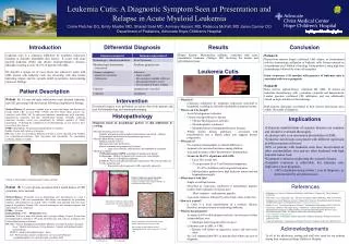
Chapter 9 Oral Manifestations of Systemic Diseases. Outline Endocrine Disorders Blood Disorders Immunodeficiency Oral Manifestations of Therapy for Oral Cancer Effects of Drugs on the Oral Cavity Oral Manifestations of Systemic Diseases. Oral Manifestations of Systemic Diseases.

E N D
Chapter 9 Oral Manifestations of Systemic Diseases
Outline Endocrine Disorders Blood Disorders Immunodeficiency Oral Manifestations of Therapy for Oral Cancer Effects of Drugs on the Oral Cavity Oral Manifestations of Systemic Diseases
Oral Manifestations of Systemic Diseases (pg. 288) Many systemic diseases are reflected in the oral mucosa, maxilla, and mandible. Mucosal changes may include ulceration or mucosal bleeding. Immunodeficiency can lead to opportunistic diseases such as infection and neoplasia. Bone disease can affect the maxilla and mandible. Systemic disease can cause dental and periodontal changes. Drugs prescribed for a systemic disease can affect oral tissue.
Oral Manifestations of Systemic Diseases (cont.) Local factors may be involved in the manifestation of systemic disease in oral mucosa. The mucosa may be more easily injured due to a systemic disease, and mild irritation and chronic inflammation may cause lesions that otherwise would not occur. These may include Endocrine disorders, disorders of red and white blood cells, disorders of platelets and other bleeding and clotting disorders, and immunodeficiency disorders
Endocrine Disorders Hyperpituitarism Hyperthyroidism Hypothyroidism Hyperparathyroidism Diabetes Mellitus Addison Disease
Endocrine Disorders (pg. 288) The endocrine system consists of a group of integrated glands and cells that secrete hormones. The secretion is controlled by feedback mechanisms. The amount of hormone circulating in blood triggers factors that control production. Diseases may result from conditions where too much or too little hormone is produced.
Hyperpituitarism (pgs. 288-289) Excess hormone production by the anterior pituitary gland Caused most often by a benign tumor (pituitary adenoma) that produces growth hormone Giantism results if it occurs before the closure of long bones. Acromegaly results when hypersecretion occurs during adult life.
Clinical Features and Oral Manifestations of Hyperpituitarism (pgs. 288-289) Affects both men and women, most commonly during the fourth decade of life Patients experience poor vision, light sensitivity, enlargement of hands and feet, and an increase in rib size. Facial changes Enlargement of maxilla and mandible may cause separation of teeth and malocclusion. Frontal bossing and an enlargement of nasal bones may lead to deepening of voice. Mucosal changes May have thickened lips and macroglossia
Clinical Features and Oral Manifestations of Hyperpituitarism (cont.)
Diagnosis and Treatment of Hyperpituitarism Diagnosis involves measurement of growth hormone. Treatment often includes pituitary gland surgery.
Hyperthyroidism (Thyrotoxicosis) (pg. 289) Excess production of thyroid hormone More common in women than men The most common cause is Graves disease Graves disease Appears to be due to an autoimmune disorder in which a substance is produced that abnormally stimulates the thyroid gland Other causes include hyperplasia of the gland, benign and malignant tumors of the thyroid, pituitary gland disease, and metastatic tumors.
Clinical Features of Hyperthyroidism Rosy complexion, erythema of the palms, excessive sweating, fine hair, softened nails The patient may have exophthalmos. Anxiety, weakness, restlessness, and cardiac problems may also be associated.
Oral Manifestations of Hyperthyroidism May lead to premature exfoliation of deciduous teeth in children and premature eruption of permanent teeth Osteoporosis may affect alveolar bone. Caries and periodontal disease may appear and develop more rapidly in these patients. Burning tongue also has been reported.
Treatment of Hyperthyroidism May include surgery, medications to suppress thyroid activity, or administration of radioactive iodine
Hypothyroidism (pg. 289) A decreased output of thyroid hormone Causes include developmental disturbances, autoimmune disease, iodine deficiency, drugs, and pituitary disease Cretinism When it occurs in infancy and childhood Myxedema When it occurs in older children and adults
Hypothyroidism (cont.) Oral manifestations In infants Thickened lips, enlarged tongue, and delayed eruption of teeth In adults Enlarged tongue
Hyperparathyroidism (pgs. 289-290) Due to excessive secretion of parathyroid hormone from the parathyroid glands The four parathyroid glands are located near the thyroid gland. Parathyroid hormone plays a role in calcium and phosphorous metabolism. Hyperparathyroidism is characterized by elevated blood levels of calcium (hypercalcemia) and low levels of blood phosphorous (hypophosphatemia).
Hyperparathyroidism (cont.) May be the result of hyperplasia of parathyroid glands, a benign tumor of one or more parathyroid glands, or a malignant parathyroid tumor Found in middle-aged adults Much more common in women than men Parathyroid hormone increases the uptake of dietary calcium from the gastrointestinal tract and is able to move calcium from bone to circulating blood when necessary.
Clinical Featuresof Hyperparathyroidism Mild cases may be asymptomatic, or may have joint pain or stiffness. Lethargy and coma may occur with severe disease.
Oral Manifestations of Hyperparathyroidism Well-defined unilocular or multilocular radiolucencies Microscopically, they appear to be CGCG (central giant cell granulomas). Bone may have a mottled appearance.
Diagnosis and Treatmentof Hyperparathyroidism Measurement of parathyroid hormone blood levels May include serum calcium and phosphorous measurements Treatment is directed at correcting the cause of increased hormone production. Causes may include tumors, renal disease, and vitamin D deficiency.
Diabetes Mellitus (pgs. 290-294) A chronic disorder of carbohydrate metabolism characterized by abnormally high blood glucose levels These result from a lack of insulin, defective insulin that does not work to lower blood glucose levels, or increased insulin resistance due to obesity.
Diabetes Mellitus (cont.) Glucose normally signals beta cells of the pancreas to make insulin. The hormone is then secreted into the bloodstream to facilitate the uptake of glucose into fat and skeletal muscle. In the presence of insulin, fat and skeletal muscle cells can use glucose as an energy source.
Diabetes Mellitus (cont.) Without insulin, tissue is broken down to provide energy and weight loss occurs. A severe hyperglycemia can lead to diabetic coma. Ketone can be produced by the breakdown of fatty acids. Ketoacidosis lowers the pH of blood. Phagocytic activity of macrophages is reduced and neutrophil chemotaxis is delayed. Collagen production is abnormal.
Types of Diabetes Insulin-dependent diabetes mellitus Type 1 Non–insulin-dependent diabetes mellitus Type 2
Insulin-Dependent Diabetes Mellitus (pgs. 291-292) Thought to be an autoimmune disease Insulin-producing cells of the pancreas are destroyed. 3% to 5% of all diabetic patients have this type. Can occur at any age, the peak is at 20 Acute onset with polydipsia (excessive thirst and intake of fluid), polyuria (excessive urination), and polyphagia (excessive appetite)
Insulin-Dependent Diabetes Mellitus (cont.) These patients will require insulin their entire lives. The current approach to management of these patients involves multiple insulin injections and proper diet, exercise, and frequent determination of blood glucose levels. But multiple injections of insulin can more readily lead to low blood sugar (hypoglycemia) and insulin shock (severe hypoglycemia).
Insulin-Dependent Diabetes Mellitus (cont.) New methods of treatment Nasal spray rather than injection Insulin pump A backup may be necessary in case the pump fails Low insulin can lead to ketoacidosis, resulting in nausea, abdominal cramps, disorientation, and fatigue
Non–Insulin-Dependent Diabetes Mellitus (pgs. 292-294) Characterized by insulin resistance 95% of all diabetic patients have this type of diabetes. Usually occurs in patients 35 to 40 years of age or older Many of these individuals are obese Obesity probably decreases the number of receptors for insulin binding in sensitive tissues like fat or muscle. Diet and weight reduction may control it in some individuals; others require oral hypoglycemic agents.
Clinical Features of Non–Insulin-Dependent Diabetes Mellitus (pg. 293) Atherosclerosis, a thickening of the blood vessel wall from fibrofatty plaques, can lead to impaired circulation, causing impaired oxygenation and nutrition in tissue. This increases the risk of ulceration and gangrene of the feet, high blood pressure, kidney failure, and stroke. Diabetic retinopathy in the eye can lead to blindness. The nervous system may be affected. The person may have decreased resistance to infection.
Clinical Features of Non–Insulin-Dependent Diabetes Mellitus (cont.)
Oral Complications of Non–Insulin-Dependent Diabetes Mellitus (pgs. 293-294) Patients may have an increased prevalence of oral candidiasis. Mucormycosis, a rare oral fungal infection that affects the palate and maxillary sinuses, may be seen in uncontrolled or poorly controlled diabetes. Bilateral asymptomatic parotid gland enlargement may occur. Xerostomia may be associated with uncontrolled diabetes mellitus. Patients may have an accentuated response to plaque. Patients may have slow wound healing and increased susceptibility to infection.
Oral Complications of Non–Insulin-Dependent Diabetes Mellitus (cont.)
Addison Disease (pg. 294) Primary adrenal cortical insufficiency In most cases, the cause of destruction of the adrenal gland is unknown – it may be an autoimmune disease. It may be due to a tumor or tuberculosis. To compensate, the pituitary gland increases production of ACTH.
Addison Disease (cont.) Clinical features This hormone causes stimulation of melanocytes. Bronzing of the skin may occur, as well as melanotic macules on oral mucosa. Treatment Steroid replacement therapy
Blood Disorders Disorders of Red Blood Cells and Hemoglobin Disorders of White Blood Cells Bleeding Disorders
Blood Disorders (cont.) (pg. 294) (Box 9-1) The complete blood count examines red blood cells, white blood cells, and platelets. It provides information about the number of each type of cell, the ratio of types, and the appearance of the cells.
Disorders of Red Blood Cells and Hemoglobin Anemia Iron Deficiency Anemia Pernicious Anemia Folic Acid and Vitamin B12 Deficiency Anemia Thalassemia Sickle Cell Anemia Celiac Sprue Aplastic Anemia Polycythemia
Anemia (pgs. 294-295) A reduction in the oxygen-carrying capacity of blood Most often related to a decrease in the number of circulating red blood cells Nutritional anemias A deficiency in a substance required for the normal development of red blood cells, commonly vitamins Suppression of bone marrow stem cells
Anemia (cont.) Clinical features Pallor of skin and oral mucosa Angular cheilitis Erythema and atrophy of oral mucosa Loss of filiform and fungiform papillae on the dorsum of the tongue
Iron Deficiency Anemia (pg. 295) An insufficient amount of iron is supplied to bone marrow for red blood cell development. May occur as a result of deficient iron intake, blood loss from heavy menstrual bleeding or chronic gastrointestinal bleeding, poor iron absorption, or an increased requirement for iron in situations such as pregnancy or infancy Plummer-Vinson syndrome may result from long standing iron deficiency anemia. Includes dysphagia, atrophy of the upper alimentary tract, and a predisposition to developing oral cancer
Clinical Features and Oral Manifestations of Iron Deficiency Anemia (pg. 295) Often asymptomatic, may have nonspecific symptoms such as weakness and fatigue In severe cases may see angular cheilitis, pallor of oral tissue, and an erythematous, smooth, painful tongue
Clinical Features and Oral Manifestations of Iron Deficiency Anemia (cont.)
Diagnosis and Treatment of Iron Deficiency Anemia Laboratory tests show a low hemoglobin content and reduced hematocrit. Red blood cells appear smaller than normal (microcytic) and light in color (hypochromic) Treatment Dietary supplements
Pernicious Anemia (pgs. 295-296) Probably an autoimmune disorder in most situations May be caused by removal of the stomach, gastric cancer, or gastritis Caused by a deficiency in intrinsic factor Intrinsic factor is secreted by parietal cells in the stomach; it is necessary for absorption of vitamin B12
Clinical Features and Oral Manifestations of Pernicious Anemia (pgs. 295-296) Weakness, pallor, and fatigue on exertion May see nausea, dizziness, diarrhea, abdominal pain, loss of appetite, and weight loss Angular cheilitis, mucosal pallor, painful atrophic and erythematous mucosa, mucosal ulceration, loss of papillae on the dorsum of the tongue, and burning and painful tongue
Clinical Features and Oral Manifestations of Pernicious Anemia (cont.)
Diagnosis and Treatment of Pernicious Anemia Laboratory tests reveal low serum B12 levels and gastric achlorhydria (lack of hydrochloric acid) Red blood cells have megaloblastic anemia. Abnormally large and immature megaloblasts with nuclei The Schilling test detects an inability to absorb oral vitamin B12 Treatment Injections of vitamin B12