Download
1 / 48
600 likes | 1.19k Vues
Chapter 9 Membrane Transport. Outline. How does transport occur across biological membranes work? What is passive diffusion? How does facilitated diffusion occur? How does energy input drive active transport processes? How are certain transport processes driven by light energy?

E N D
Outline • How does transport occur across biological membranes work? • What is passive diffusion? • How does facilitated diffusion occur? • How does energy input drive active transport processes? • How are certain transport processes driven by light energy? • How is secondary active transport driven by ion gradients?
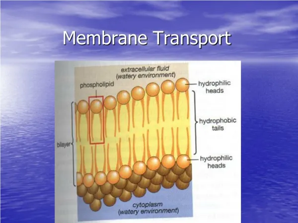
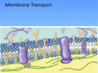
9.5 How Does Transport Occur Across Biological Membranes? • Transport processes are vital for all life forms • Cells must be able to import nutrients and export waste • All cells maintain concentration gradients of various metabolites across the plasma membrane and other intracellular membranes • Many transport processes involve movement of a polar, water-soluble molecule or ion across the hydrophobic interior of a membrane • Specific transport proteins accomplish these tasks • Transport proteins are integral membrane proteins
9.6 What is Passive Diffusion? No special proteins needed • Transported species simply moves down its concentration gradient - from high [c] to low [c] • High permeability coefficients usually mean that passive diffusion is not the whole story
9.6 What is Passive Diffusion? Passive diffusion of an uncharged species across a membrane depends only on the concentrations on the two sides.
9.6 What is Passive Diffusion? Passive diffusion of a charged species depends on concentration and also on the charge of the particle and the electrical potential difference across the membrane.
9.7 How Does Facilitated Diffusion Occur? ΔG negative, but proteins assist • Solutes only move in the thermodynamically favored direction • But proteins may "facilitate" transport, increasing the rates of transport • Two important distinguishing features: • solute flows only in the favored direction • transport displays saturation kinetics
9.7 How Does Facilitated Diffusion Occur? Passive diffusion and facilitated diffusion may be distinguished graphically. The plots for facilitated diffusion are similar to plots of enzyme-catalyzed processes, and they display saturation behavior.
9.7 How Does Facilitated Diffusion Occur? Passive diffusion and facilitated diffusion may be distinguished graphically. The plots for facilitated diffusion are similar to plots of enzyme-catalyzed processes, and they display saturation behavior.
9.7 How Does Facilitated Diffusion Occur? Passive diffusion and facilitated diffusion may be distinguished graphically. The plots for facilitated diffusion are similar to plots of enzyme-catalyzed processes, and they display saturation behavior.
Membrane Channels Can be Formed in a Variety of Ways Single channel pores can be formed from dimers, trimers, tetramers or pentamers of protein subunits. Multimeric assemblies in which each subunit has its own pore are known.
Potassium Channels Combine High Selectivity with High Conduction Rates Structure of the KcsA potassium channel from S. lividans. The four identical subunits of the channel, which surround a central pore, are shown in different colors.
Potassium Channels Combine High Selectivity with High Conduction Rates Structure of the KcsA potassium channel from S. lividans. Each subunit contributes three α–helices (blue, green, red) to the tetrameric structure.
Potassium Channels Combine High Selectivity with High Conduction Rates Structure of the KcsA potassium channel from S. lividans. The selectivity filter is made from loops from each of the subunits. The four primary K+ binding sites are numbered.
Potassium Channels Combine High Selectivity with High Conduction Rates Structure of the KcsA potassium channel from S. lividans. The tetrameric channel, as viewed through the pore.
Potassium Channels Combine High Selectivity with High Conduction Rates Model for outward and inward transport through the KcsA potassium channel. The selectivity filter in the channel contains four K+-binding sites, only two of which are filled at any time.
The KcsA channel is gated by intracellular pH. It is closed at neutral pH, open at low pH. Helix bending and rearrangement deep in the membrane opens K+ channels. The closed and open states of the potassium channel.
Gating in voltage-dependent K+ channels Model for voltage-dependent gating in mammalian K+ channels. Parts a and c show a single subunit of the channel. Parts c and d show the native tetrameric channel. Parts a and c are the open channel conformation. Parts b and d show the closed state. (pdb id = 1J4C. Photo courtesy of Roderick MacKinnon.
Selectivity Filters Made From Peptide Loops Control Ion Channel Selectivity Comparison of selectivity filters for a variety of channels shows that variations in sequence change the selection for ions. Ion selectivity in cation channels is a function of peptide sequence of the selectivity filter. Conserved Gly residues are red. Similar amino acids are yellow.
The NaK channel of B. cereus is similar to K+ channels, but with equal affinity for Na+ and K+ The selectivity filter sequence for this channel is TVGDG. Structure of the channel from Bacillus cereus, which has equal preference for Na+ and K+. (a) The front subunit has been removed to show the 5 ion-binding sites. (b) A view of the channel through the pore. (c) Substitution of D for Y in the filter eliminates sites 1 and 2, leaving a pore vestibule that bind K+ with low affinity.
Chloride, Water, Glycerol, and Ammonia Flow Through Single-Subunit Pores Structures of the channels for (a) chloride, (b) ammonia, and (c) glycerol. All are axial views.
9.8 How Does Energy Input Drive Active Transport Processes? Energy input drives transport • Some transport must occur such that solutes flow against their thermodynamic potential • Energy input drives such transport • Energy source and transport machinery are "coupled" • Energy source may be ATP, light or a concentration gradient
The Sodium Pump aka Na,K-ATPase • Large protein - 120 kD and 35 kD subunits • Maintains intracellular Na+ low and K+ high • Crucial for all organs, but especially for neural tissue and the brain • ATP hydrolysis drives Na+ out and K+ in • Alpha subunit has ten transmembrane helices with large cytoplasmic domain
Na+,K+-ATPase Uses ATP Energy to Drive Sodium and Potassium Transport
Na+,K+-ATPase Uses ATP Energy to Drive Sodium and Potassium Transport • ATP hydrolysis occurs via an E-P intermediate • Mechanism involves two enzyme conformations known as E1 and E2 • Cardiac glycosides inhibit by binding to outside
Na+,K+-ATPase Uses ATP Energy to Drive Sodium and Potassium Transport A mechanism for Na+,K+-ATPase. The model assumes two principal conformations, E1 and E2. Binding of Na+ ions to E1 is followed by phosphorylation and release of ADP.
Calcium Transport Is Accomplished in the Sarcoplasmic Reticulum by Ca2+-ATPase A process akin to Na,K transport • Calcium levels in resting muscle cytoplasm are maintained low by Ca2+-ATPase - a Ca2+ pump • Calcium is pumped into the sarcoplasmic reticulum (SR) by a 110 kD protein that is very similar to the alpha subunit of Na,K-ATPase • Aspartyl phosphate E-P intermediate is at Asp-351 and Ca2+-pump also fits the E1-E2 model
Calcium Transport Is Accomplished in the Sarcoplasmic Reticulum by Ca2+-ATPase The transport cycle of the sarcoplasmic reticulum Ca2+-ATPase involves at least five different conformations of the protein, represented by the blue-shaded boxes here, and shown on the following slides.
The Gastric H+,K+-ATPase The enzyme that keeps the stomach at pH 0.8 • The parietal cells of the gastric mucosa (lining of the stomach) have an internal pH of 7.4 • H+,K+-ATPase pumps protons from these cells into the stomach to maintain a pH difference across a single plasma membrane of 6.6! • This is the largest known transmembrane gradient in eukaryotic cells
The Gastric H+,K+ -ATPase Maintains the Low pH of the Stomach The H+,K+-ATPase and a K+/Cl− cotransport system work together to achieve net transport of H+ and Cl−.
The Gastric H+,K+-ATPase • H+,K+-ATPase is similar in many respects to Na+,K+-ATPase and Ca2+-ATPase • All three enzymes form covalent E-P intermediates • All three have similar sequences for the large (α) subunit
Bone Remodeling by Osteoclast Proton Pumps How your body takes your bones apart: • Bone material undergoes ongoing remodeling • osteoclasts tear down bone tissue • osteoblasts build it back up • Osteoclasts function by secreting acid into the space between the osteoclast membrane and the bone surface - acid dissolves the Ca2+-phosphate matrix of the bone • An ATP-driven proton pump in the membrane does this
Bone Remodeling by Osteoclast Proton Pumps • Vacuolar (V-type) ATPases are found in vacuoles, lysosomes, endosomes and other organelles • A vacuolar ATPase is present in osteoclasts (multinucleate cells that break down bone during normal bone remodeling) • This osteoclast ATPase secretes acid (protons) into the space between the bone surface and the cell • This transport of protons out of the osteoclast lowers the pH of the extracellular space near the bone to about 4, dissolving the crystalline hydroxyapatite lattice of the bone
Bone Remodeling by Osteoclast Proton Pumps Proton pumps cluster in the ruffled border of osteoclasts and pump protons into the space between the cell and the bone.
ABC Transporters Use ATP to Drive Import and Export Functions and Multidrug Resistance • Cells “clean house” with membrane transporters known as multidrug resistance (MDR) pumps • MDR pumps are designed to recognize foreign organic molecules in cells and pump them out • In bacteria, these pumps use the hydrolytic energy of ATP to import nutrients into the cell • At least five families of influx and efflux pumps are known • Among these are the ABC transporters, which export therapeutic drugs from cancer cells
ABC Transporters Use ATP to Drive Import and Export Functions and Multidrug Resistance • All ABC transporters consist of two transmembrane domains (TMDs) which form the pore and two cytosolic nucleotide-binding domains (NBDs) that bind and hydrolyze ATP • Bacterial ABC transporters are multimeric, but eukaryotic ABC pumps are monomeric
ABC Transporters Use ATP to Drive Import and Export Functions and Multidrug Resistance Influx pumps in the inner membrane of Gram-negative bacteria bring nutrients into the cell; efflux pumps export cellular waste products.
9.9 How Are Certain Transport Processes Driven by Light Energy? Bacteriorhodopsin is a light-driven proton pump • Halobacterium halobium, the salt-loving bacterium, carries out normal respiration if O2 and substrates are plentiful • But when substrates are lacking, it can survive by using bacteriorhodopsin and halorhodopsin to capture light energy • Purple patches of H. halobium are 75% bR and 25% lipid - a "2D crystal" of bR - ideal for structural studies
9.9 How Are Certain Transport Processes Driven by Light Energy? Protein opsin and retinal chromophore • Retinal is bound to opsin via a Schiff base linkage • The Schiff base (at Lys216) can be protonated, and this site is one of the sites that participate in H+ transport • The carboxyl groups of Asp85 and Asp96 also serve as proton binding sites during transport • These Asp residues lie in hydrophobic environments; their carboxyl pKa values are near 11.
9.9 How Are Certain Transport Processes Driven by Light Energy? The Schiff base linkage between the retinal chromophore and Lys216.
9.9 How Are Certain Transport Processes Driven by Light Energy? • Lys216 is buried in the middle of the 7-TMS structure of bR, and retinal lies mostly parallel to the membrane and between the helices • Light absorption converts retinal from all-trans to 13-cis configuration, triggering conformation changes that induce pKa changes • This facilitates proton transfers from Asp96 to the Lys Schiff base to Asp85 and net proton transport across the membrane
A Proton Transport Model The mechanism of proton transport by bacteriorhodopsin. The hydrophobic environments of Asp85 and Asp96 raise the pKa values of their side chain carboxyl groups, making it possible for these carboxyls to accept protons as they are transported across the membrane.
9.10 How Is Secondary Active Transport Driven by Ion Gradients? • The gradients of H+, Na+ and other cations and anions established by ATPases can be used for secondary active transport of various substrates • Many amino acids and sugars are accumulated by cells in transport processes driven by Na+ and H+ gradients • Many of these are symports, with the ion and the transported amino acid or sugar moving in the same direction • In antiport processes, the ion and the transported species move in opposite directions
AcrB is a Secondary Transport System • AcrB is the major MDR transporter in E. coli • It is responsible for pumping a variety of molecules • AcrB is part of a tripartite complex that bridges the E. coli inner and outer membranes and spans the entire periplasmic space • AcrB works with AcrA and TolC to transport drugs and other toxins from the cytoplasm across the entire cell envelope and into the extracellular medium
AcrB is a Secondary Transport System A tripartite complex of proteins comprises the large structure in E. coli that exports waste and toxin molecules. The transport pump is AcrB, embedded in the inner membrane.
AcrB is a Secondary Transport System • AcrB is a secondary active transport system and a H+-drug antiporter • As protons flow spontaneously inward through AcrB in the E. coli inner membrane, drug molecules are driven outward • Remarkably, the three identical subunits of AcrB adopt slightly different conformations, denoted loose (L), tight (T), and open (O) • These three conformations are three consecutive states of a transport cycle • As each monomer cycles through L, T, and O states, drugs enter tunnel, are bound and then exported
AcrB is a Secondary Transport System A model for drug transport by AcrB involves three different conformations.