Download
1 / 14
150 likes | 229 Vues
Learn about OCT technology, how it works, its applications in ophthalmology, and the principles behind its use in imaging biological tissues. Explore the setup, resolution factors, and exemplary models.

E N D
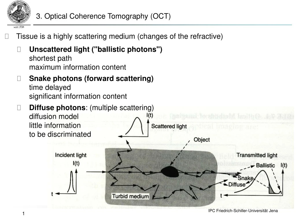
3. Optical Coherence Tomography (OCT) • Tissue is a highly scattering medium (changes of the refractive) • Unscattered light ("ballistic photons") shortest pathmaximum information content • Snake photons(forward scattering)time delayedsignificant information content • Diffuse photons: (multiple scattering) diffusion modellittle information to be discriminated
3. Optical Coherence Tomography (OCT) • Identifies scatterers by interference with incoherent reference (Michelson interferometer) • Reference beam interferes with ballistic photons from scattering sample • Fully coherent source no selectivity to photons from a specific depth • White light: Interference only when path difference is within coherence length(a specific depth in sample) • By scanning the reference mirror a depth discrimination is achieved
The OCTsetup Fiber-optic beamsplitter Broadband source Tissue Scanning reference mirror Detector Computer Amplifier Bandpass filter
Interference Coherent source Michelson interferometer light source Detector Partially coherent source
3. Optical Coherence Tomography (OCT) Exemplary model for a sample comprising a series of discrete reflectors. Andrew Gomez, Daniel Kim, Jiwon Lee, Kenny Tao Izatt, Joseph A. Theory of Optical Tomography, 2006 http://www.duke.edu/~yt13/Optical%20Coherence%20Tomography.ppt
3. Optical Coherence Tomography (OCT) k=2p/l 0 zS-zR
3. Optical Coherence Tomography (OCT) • Axial resolution Dz is determined by coherence length DL of the light source i.e. the shorter the coherence length the better the axial resolution • Application of a broad band light source e.g. super-luminescent diode, photonic bandgap fibers • Lateral resolution is determined by the diffraction limited spot size of the focus • A-Scan: assigns every investigated depth point a certain reflectivity • B-Scan: reassembling of multiple A-scans by laterally scanning the light beam along a line • C-Scan: three-dimensional tomography by laterally scanning in two dimensions l0 = center wavelength of the broad band light source Dl = width of the broad band light source (assumption: Gaussian spectrum)
Reference beam Eye Beam splitter Light source Signal analysis Detector 3. Optical Coherence Tomography (OCT) • Clinical application of OCT in Ophthalmology In vivo OCT scan of a retina @ 800 nm (axial resolution = 3 µm) Cornea OCT image
Resolution (log) 1 mm Ultrasound 100 mm 10 mm Confocalmicroscopy 1 mm Penetration depth (log) 1 mm 1 cm 10 cm OCT vs. standard imaging Standardclinical Highfrequency OCT from: Peter E. Anderson, DTU course 2004
3. Optical Coherence Tomography (OCT) • Curvature of OTFs • Use extended focus techniques? • Problem: • HF information is translated to low frequencies (wrong)
4. Molecular many electron systems: electronic & nuclear movement
4. Molecular many electron systems: electronic & nuclear movement • Hamiltonian for a polyatomic molecule treated as Coulomb system with N nuclei (coordinates {R}) and n electrons (coordinates {ri}) : In atomic units i.e. ~= qe = me = 1 Kinetic energy operator for nuclei Kinetic energy operator for electrons Nuclei-electron interaction operator Electron-electron interaction operator Nuclei-nuclei interaction operator
4. Molecular many electron systems: electronic & nuclear movement • (3N + 3n)-dimensional problem:Born-Oppenheimer Approximation: separate treatment of electronic and nuclear motion allows the total wavefunction of a molecule to be broken into its electronic and nuclear components: Does not depend on {ri} = constant for given nuclear geometry Decomposition of Hamiltonian: = adiabatic potential energy surfaces Schrödinger equation for complete problem: