Download
1 / 24
240 likes | 1.37k Vues
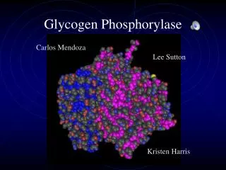
Carlos Mendoza. Lee Sutton. Glycogen Phosphorylase. Kristen Harris. Glycogen Phosphorylase is an enzyme found in glycogen granules in the cytosol

E N D
Carlos Mendoza Lee Sutton Glycogen Phosphorylase Kristen Harris
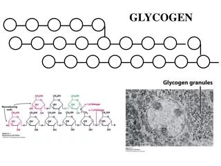
Glycogen Phosphorylase is an enzyme found in glycogen granules in the cytosol • Glycogen Phosphorylase releases sugar from glycogen by clipping off glucose from the chains of a glycogen molecule. It catalyzes a phosphorolysis reaction where it adds a phosphate to break off glucose: (glucose)n + Pi <<====>> glucose 1-phosphate + (glucose)n-1
Protein Characteristics 1 kqisvrglag venvtelkkn fnrhlhftlv kdrnvatprd yyfalahtvr dhlvgrwirt 61 qqhyyekdpk riyylslefy mgrtlqntmv nlalenacde atyqlgldme eleeieedag 121 lgngglgrla acfldsmatl glaaygygir yefgifnqki cggwqmeead dwlrygnpwe 181 karpeftlpv hfygrvehts qgakwvdtqv vlampydtpv pgyrnnvvnt mrlwsakapn 241 dfnlkdfnvg gyiqavldrn laenisrvly pndnffegke lrlkqeyfvv aatlqdiirr 301 fksskfgcrd pvrtnfdafp dkvaiqlndt hpslaipelm rvlvdlerld wdkawevtvk 361 tcaytnhtvi pealerwpvh lletllprhl qiiyeinqrf lnrvaaafpg dvdrlrrmsl 421 veegavkrin mahlciagsh avngvarihs eilkktifkd fyelephkfq nktngitprr 481 wlvlcnpgla eiiaerigee yisdldqlrk llsyvddeaf irdvakvkqe nklkfaayle 541 reykvhinpn slfdvqvkri heykrqllnc lhvitlynri kkepnkfvvp rtvmiggkaa 601 pgyhmakmii klitaigdvv nhdpvvgdrl rviflenyrv slaekvipaa dlseqistag 661 teasgtgnmk fmlngaltig tmdganvema eeageenffi fgmrvedvdr ldqrgynaqe 721 yydripelrq iieqlssgff spkqpdlfkd ivnmlmhhdr fkvfadyeey vkcqervsal 781 yknprewtrm virniatsgk fssdrtiaqy areiwgveps rqrlpapdea ap • sizeof(Glycogen Phosphorylase) = 832 aa 8GPB Amino acid chain In humans found on chromosome 20
Glycogen Phosphorylase can be in a dimer or tetramer form. However, it is thought to be mostly in the dimer form in vivo. Each monomer consists of two domains: • C terminal domain - domain responsible for catalysis of the reaction (blue) • N terminal domain - domain responsible for regulation of the enzyme (pink)
Both domains consist of a beta sheet core surrounded by layers of alpha helical segments
C TERMINAL DOMAIN • The active site is actually in the cleft between the C and N terminal domains • A cofactor PLP (pyridoxal phosphate) binds near the active site to facilitate the reaction
N TERMINAL DOMAIN • Contains different allosteric effector sites that determine when the enzyme is active (R state) or inactive (T state.) • Can be phosphorylated at Serine 14 to induce active state (see previous slide) • Or can bind with Amp to induce active state
Change from T to R state • Problem - T state is found as a monomer, but R state only crystallized as a tetramer. • Glycogen Phosphorylase forms tetramers when there is a lot of the enzyme present, which is the case when glucose is needed and there is a lot of the R state of the enzyme present. • This makes the two hard to compare visually.
Upon phosphorylation - • Amino acids 10-22 swing 120 degrees causing a change in both the N and C domain overall structures
Additional change from T to R State • Residues 280-286 go from blocking active site in T state to pulled out away from it in the R state.
AMINO ACIDS 280-286 IN YELLOW R (ACTIVE) T (INACTIVE)
MAIN POINT IS THAT BINDING AT EFFECTOR SITES FOR PHOPSHATE OR AMP LEAD TO CHANGES IN THE PROTEIN’S SHAPE THAT MAKES IT MORE ABLE TO ACCEPT GLYCOGEN AT IT’S ACTIVE SITE
unrooted Phylogenetic trees
Tree characteristics All sequences are closely conserved Bacteria tend to be more closely related to other bacteria than to the others The brain, liver, and muscle forms of Glycogen phosphorylase tend to group together The fruit fly is grouped closer to the vertebrates than to the bacteria as expected
The brain, liver, and muscle forms in one organism are almost identical to the brain, liver, and muscle forms in different organisms Glycogen phosphorylase showed up in a lot of bacteria, and then in vertebrates, but not much in the middle this might be because it hasn’t been studied much in other organisms
Evolution of Glycogen phosphorylase Over time glycogen phosphorylase has changed very little it is very similar in bacteria and vertebrates There is more variation in bacteria glycogen phosphorylase than in vertebrates This might be to the fact that bacteria has been around much longer and has had more time to change Or it might be that Glycogen phosphorylase in vertebrates is unable to change with out causing major problems changes in bacteria are not as damaging
Hydrophobicity • Exist in a hydrophilic environment • The percentage of solvent-accessible hydrophobic elements is much greater than in the “average” globular protein
Grease PDB entry 8GPB
Blast Not much was returned besides glycogen phosphorylase A few other enzymes that break down sugars were returned such as maltodextrin phosphorylase and Glucan phosphorylase
Software Tools • Structure Visualization : Cn3D software, using images from NCBI. • Phylogenetic Analysis : Used ClustalW on the Biology Workbench • Hydrophobicity Analysis: Used Grease on the Biology Workbench • BlastP used in biology Workbench to search for similar proteins • Used Google for everything
References Fletterick, Robert and Stephen Sprang. Glycogen phosphorylase Structures and Function.Department of Biochemistry and Biophysics, School of Medicine, University of California. Acc. Chem Res. 1982, 15, 361-369. Goodsell, David. Glycogen Phosphorylase. Molecule of the Month. Protein Data Bank. Internet. http://www.rcsb.org/pdb/molecules/pdb24_1.html. 5/13/03. Gillis, Jeremy. Glycogen Phosphorylase - Structure and Function Term Paper. Internet. http://www.chem.uwec.edu/webpapers_F98/gillis/Structure/struct_descrip.html. 5/13/03.