Management of Empyema Thoracis, Lung Abscess, and Bronchiectasis in Adult Patients
150 likes | 1.21k Vues
This report details the clinical journey of a 34-year-old female with pneumococcal pneumonia and empyema requiring intensive treatment, including tube drainage, ventilation, tracheostomy, and decortication. Follow-up imaging showed significant improvement. Additionally, we present the case of an elderly female with a lung abscess linked to a pharyngeal pouch, treated for aspiration pneumonia without organism identification. The bronchiectasis case highlights the characteristic radiological findings seen in patients with cystic fibrosis, emphasizing the importance of CT imaging in these diagnoses.

Management of Empyema Thoracis, Lung Abscess, and Bronchiectasis in Adult Patients
E N D
Presentation Transcript
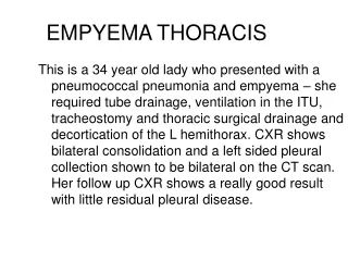
EMPYEMA THORACIS This is a 34 year old lady who presented with a pneumococcal pneumonia and empyema – she required tube drainage, ventilation in the ITU, tracheostomy and thoracic surgical drainage and decortication of the L hemithorax. CXR shows bilateral consolidation and a left sided pleural collection shown to be bilateral on the CT scan. Her follow up CXR shows a really good result with little residual pleural disease.
female DOB 1977 2009 November
LUNG ABSCESS • This elderly lady has an abscess in the apical segment of her L lower lobe. She complained of food regurgitation and was shown to have a pharyngeal pouch. No organism was identified, she was treated for aspitation pneumonia with amoxycillin and metronidazole and drainage of the abscess aided by the physiotherapists. She was deemed too frail to have the pouch repaired.
BRONCHIECTASIS • Bronchiectasis is a radiological diagnosis, bronchi are larger than normal and may have and uneven mucosa; the first CT scan shows a good example of signet ring brochovascular images –a bronchus and it’s accompanying vessel should be of similar size, where the bronchus is larger a signet ring appearance is seen. The next CT cut shows gross brochiectasis in a patient with cystic fibrosis.