STREPTOCOCCUS
STREPTOCOCCUS. Pavithra G. Palan. Streptococci are Gram positive cocci. Arranged in chains or pairs. The name streptococci ( streptos meaning twisted or coiled) was given by Billroth. 0 2 requirement. CLASSIFICATION :. Haemolysis. Serological Grouping (C carbohydrate antigen).

STREPTOCOCCUS
E N D
Presentation Transcript
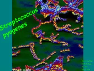
STREPTOCOCCUS Pavithra G. Palan
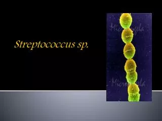
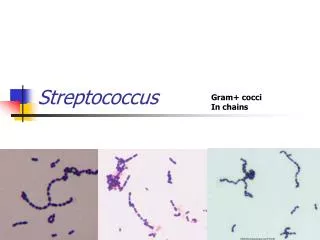
Streptococci are Gram positive cocci. • Arranged in chains or pairs. • The name streptococci (streptos meaning twisted or coiled) was given by Billroth.
02requirement CLASSIFICATION: Haemolysis Serological Grouping (C carbohydrate antigen) Group A- Streptococcus pyogens Serological typing (M Protein)
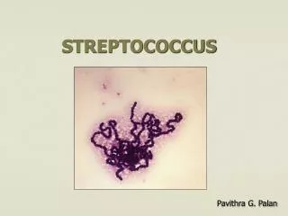
MORPHOLOGY: Gram positive spherical or oval cocci arranged in chains. Individual coccus will be 0.5-1.0μm in diameter. They are nonmotile and nonsporing. Some strains have capsule composed of hyaluronic acid. Streptococcus pyogens
CULTURE: Media used: 1. Non selective media:- Sheep blood agar 2. Selective media:- Crystal violet blood agar PNF medium
Cultural characteristics: On blood agar, after overnight incubation, the colonies are small, circular, low convex with an area of β-haemolysis around them.
Biochemical reactions: 1. Catalase test: Negative
2. Bile solubility test: Negative Negative Positive
3. PYR test: Positive 4. Ribose is not fermented.
PATHOGENICITY: Source of infection: 1. Patient 2. Carriers Mode of transmission: 1. Contact: direct or indirect( through fomites) 2. Inhalation of air borne droplets
Antigenic structure: 1.Capsular hyaluronic acid: 2.Cell wall antigens: a) Inner layer of peptidoglycan b) Middle layer of group specific C carbohydrate c) Outer layer of protein (fimbriae) & lipoteichoic acid 3.Type specific antigens: a) M protein b) T protein c) R protein
Virulence factors: These include A) Toxins B) Enzymes
A) Toxins: 1. Haemolysins a) Streptolysin ‘O’ b) Streptolysin ‘S’ 2. Pyrogenic exotoxin( Erythrogenic toxin)
B) Enzymes: 1. Streptokinase (Fibrinolysin) 2. Deoxyribonuclease (Streptodornase) 3. NADase 4. Hyaluronidase
Diseases: Diseases caused by S. pyogens is studied under 2 groups 1. Suppurative infections 2. Non suppurative complications
1. Suppurative infections: • Pyogenic infections. • Spreads locally, along lymphatics and through the blood stream.
Common suppurative infections are: A) Respiratory infections:- • Tonsillitis sore throat • Pharyngitis • Otitis media • Mastoiditis • Quinsy • Ludwig’s angina • Rarely it may cause pneumonia & meningitis • Scarlet fever (sore throat & skin rash)
Tonsillitis Pharyngitis
Mastoiditis Otitis media
Quinsy Ludwig’s angina
B) Skin infections:- • Infection of wounds & burns • Impetigo • Erysipelas
Impetigo Erysipelas
C) Soft tissue infections:- i) Cellulitis
iii) Soft tissue infections with some M types of strains may sometime cause toxic shocksyndrome resembling staphylococcal TSS.
D) Genital Infections:-Puerperal sepsis E) Other suppurative infections:- • Pyemia • Septicemia • Abscesses in internal organs such as brain, lungs, liver and kidney.
2. Non suppurative complications: • It is also called as post streptococcal complications • Non suppurative complications of S.pyogens occur 1-4 weeks after the acute infection. • The organism may not be detectable when these complications set in.
These complications are believed to be the result of hypersensitivity to some streptococcal components. • The complications are- 1. Acute rheumatic fever 2. Acute glomerulonephritis
1. Acute rheumatic fever: It occurs after repeated sore throat caused by S. pyogens. Mechanism of pathogenesis: During primary infection antibodies will be produced against some streptococcal antigen. Since streptococcal antigen has similarity with cardiac tissue antigen, the antibodies will cross react with cardiac tissue antigen causing destruction. Leads to clinical symptoms such as Aschoff’s nodules, carditis, fever and malaise.
2. Acute glomerulonephritis: It follows after skin infection caused by S. pyogens nephritogenic types. Mechanism of pathogenesis: During skin infection caused by nephritogenic types of S. pyogens, the antibodies will be produced against cell membrane antigen. These antibodies cross react with glomerular basement membrane antigen causing destruction. Leads clinical symptoms such as proteinuria, haematuria & hypertension.
LABORATORY DIAGNOSIS: A) In acute suppurative infection Specimens to be collected: • Throat swab, • Pus, • Tissue material, • Blood, • Swab from nose for detection of carriers. Transport media: Pike’s medium
I) Direct Microscopy: Direct microscopy with Gram stained smear is useful in case of pus & CSF, where cocci in chains are seen. This is of no value for specimen like sputum & genital swabs where mixed flora are normally present. Methods of examination:-
c) Gram’s staining: Smears are examined from the culture plate and reveals Gram positive cocci in chains. II) Culture: a) Media used: b) Cultural Characteristics:
d) Biochemical reactions: e) Bacitracin (1 unit/ml) sensitivity test: S. pyogens is sensitive.
f) Lancefield sero grouping: Based on ‘C’ carbohydrate antigen g) Sero typing: sero typing of S. pyogens is required only for epidemiological purposes. III) Antigen detection: ELISA & Agglutination tests are used for detection of S. pyogens antigen from throat swabs
B) In Non-suppurative complications • Serological tests are useful • The tests are – 1. Anti Streptolysin O (ASO) test 2. Anti Deoxyribonuclease B (anti-DNAase B) test 3. Anti Hyaluronidase test 4. Streptozyme test
TREATMENT: • Penicillin G is the drug of choice. • In patients allergic to penicillin; erythromycin or cephalexin is used. • Antibiotics have no effect on established glomerulonephritis & rheumatic fever.
EPIDEMIOLOGY: • Streptococcal infections of the respiratory tract are more frequent in children5-8 years of age. • They are more common in winter in the temperate countries. • Crowding is an important factor in the transmission of infections. • Outbreaks of infection may occur in closed communities such as boarding school or army camps.
PROPHYLAXIS: • Prophylaxis is indicated only in the prevention of rheumatic fever. • It is done by long term administration of penicillin in children who have developed early signs of rheumatic fever. • Antibiotic prophylaxis is not useful in case of glomerulonephritis.
OTHER HAEMOLYITC STREPTOCOCCI: Group B: Streptococcus agalactiae • It is a important pathogen of cattle & causes bovine mastitis. • In human it inhabitats genital tract.
Clinical significance: 1. Infection in neonates: 2 types a. Early onset disease- Occurs during first week of life. Source of infection- Vagina of the mother & infection is acquired during birth. Clinical symptoms- Septicemia, meningitis & pneumonia.
b. Late onset disease- Occurs during 2nd & 12th week of life. Source of infection: It usually acquired from the hospital environment. Clinical symptoms- Osteomyelitis, arthritis, conjunctivitis, respiratory infection, endocarditis & peritonitis. 2. Infection in adult: It causes bacteraemia, sepsis , wound infection, septic abortion & puerperal sepsis.
Laboratory diagnosis: Specimens collected: Blood, CSF & exudates from lesions. Methods of examinations: 1. Detection of antigen in clinical samples. 2. Direct microscopy by doing Gram’s smear.
3. Culture- Blood agar is used for culture. 4. Identification- a. Small β-haemolytic colonies on blood agar b. Gram staining c. Catalase test- Negative
f. Lancefield sero grouping is done for confirmation. Treatment: Penicillin is used.