Download
1 / 31
310 likes | 439 Vues
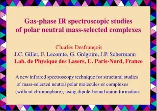
Gas-phase IR spectroscopic studies of polar neutral mass-selected complexes. A new infrared spectroscopy technique for structural studies of mass-selected neutral polar molecules or complexes (without chromophore), using dipole-bound anion formation. Charles Desfrançois

E N D
Gas-phase IR spectroscopic studiesof polar neutral mass-selected complexes A new infrared spectroscopy technique for structural studies of mass-selected neutral polar molecules or complexes (without chromophore), using dipole-bound anion formation. Charles Desfrançois J.C. Gillet, F. Lecomte, G. Grégoire, J.P. Schermann Lab. de Physique des Lasers, U. Paris-Nord, France
very diffuse orbital e- repulsion M potential energy M r ~ 10-100 Å M- potential energy distance e- - dipole m > 2 D bound state Eb ~ 0.01 eV dipolar attraction -µ/r2 -Q/4r3 -a/2r4 inter- or intra- molecular coordinate Dipole-bound anions: « neutral » ions
k (M-) Xe(nf) + M(µ) Xe+ + M-(Eb) we measure k(M-) as a function of n Pulsed dye laser 460-540 nm; n = 6-50 Collision region Molecular or cluster supersonic beam: valve + oven + carrier gas Electrostatic lens Field-detachment grids Extraction and acceleration grids Atomic Xe beam Electron gun: metastable Xe Ion detector Mass spectrum Anion time-of-flight Dipole-bound anion formation: Rydberg Electron Transfert (RET) The RET technique is selective with respect to the excess electron binding energy Eb that depends on the total dipole moment µ and thus on the neutral parent geometry.
A simple system: formamide – water complex DD0 (meV) µ (D) Ebth= 130 meV 177 6.46 113 3.94 Ebth= 30 meV isolated formamide: µ = 3.9 D; Ebth = 15 meV Ebexp = 16 meV formamide-water complex: lowest configuration µ = 2.7 D; Ebth = 2.8 meV Ebexp = 3.1 meV Ebth= 2.8 meV 0 2.69 Ebexp= 3.1 meV
Less simple: N-methylformamide - water complex Ebth= 125 meV DDe (meV) µ (D) trans NMF cis µ (D) DDe (meV) 100 6.25 4.45 111 Ebth= 29 meV Ebth= 39 meV Ebexp= 29 and3.5meV 12 4.36 0 3.24 Ebth= 4.2 meV 0 4.02 Ebth= 28 meV Need for more experimental data !
Coupling dipole-bound anion formation and IR spectroscopy • Dipole-bound anion formation by RET is a unique ionisation method that is almost totally non-perturbative: almost no internal energy is provided upon ionisation. It allows rigorous mass-selection and partial structure-selection. It is an alternative technique to REMPI when polar molecules without chromophore are involved. • If resonant IR absorption occurs, for C-H N-H or O-H bonds, the neutral molecule or complex will acquire a lot of internal energy (2800-3800 cm-1) and then: * molecular dipole-bound anions will autodetach * neutral non-covalent complexes will predissociate In both cases, anion signal will decrease.
D0 = 0.28 eV neutral vibrational predissociation Pulsed dye laser 460-540 nm; n = 6-50 hn = 0.42 eV dipole-bound anion autodetachment IR laser IR OPO laser ~ 100 µs before the dye laser Scan: 2500-4000 cm-1 Molecular or cluster supersonic beam: valve + oven + carrier gas Electrostatic lens Field-detachment grids Extraction and acceleration grids Electron gun: metastable Xe Ion detector Mass spectrum Anion time-of-flight Atomic Xe beam Rydberg Electron Transfer (RET) + IR Spectroscopy Typical anion signal: 0.1/laser shot Rydberg laser: 20 Hz; IR OPO laser: 10 Hz Real-time anion signal depletion but still shot-to-shot fluctuations
IR spectrum water dimer sym. : IR laser full bars: absolute anharmonic values dash bars: scaled harmonic values MP2 / 6-31++G(2d,2p) calculations:
IR spectrum formamide - water complex : IR laser full bars: absolute anharmonic values dash bars: scaled harmonic values MP2 / 6-31++G(2d,2p) calculations:
IR spectrum NMF – water high dipole conformers red blue full bars: absolute anharmonic values dash bars: scaled harmonic values MP2 / 6-31++G(2d,2p) calculations:
Isolated formamide and dimer F: dipole-bound µ = 3.72 D Eb = 16.5 meV F2: quadrupole-bound µ = 0 D; Q = +48 DÅ Eb = 11.3 meV D0 = 0.47 eV Dimer Formamide vibrational autodetachment is less efficient than vibrational predissociation
Ongoing work • To improve the dipole-bound anion signals: Rubidium Rydberg source (2-photon excitation). • To switch to a laser desorption source for the molecular beam. • To extend the IR OPO to the 4-10 µm region (AgGaSe2).
Calculations of glycine – water clusters : neutrals, zwitterions and anionsCharles Desfrançois, Lab. de Physique des Lasers, Université Paris 13Sungyul Lee, Kyung Hee University, Korea Goals To understand Bowen’s and Johnson’s PES results: DB anions for GWn, n=0,1,2 with Eb= 0.095, 0.195, 0.14/0.33 eV Threshold for stable (valence ?) anions: 4-5 water VDE = 0.62 eV EAad = 0.4 eV To follow the hydration transition from neutrals to zwitterions Is it 4-5 water molecules (Bowen exp.) ? Or 7-8 (Gordon calc.) To check for dipole-bound anions vs “zwitteranions” What are the dipole moments of the lowest neutrals ?
PES data 0.4 eV Gly- VDE = 95 meV EA ~ 0.4 eV VDE ~ 0.6 eV Gly-W1 VDE = 0.195 eV Gly-W2 VDE = 0.14 eV VDE = 0.33 eV
Methods Search for G(H2O)n equilibrium structures and energies For GI, GII, (GIII), GZ and GZ- and for n = 0, 1, 2, 3, 4, 5… Use of home-made force-field and genetic algorithm (MINGEN) In order to get starting structures within 0.2 eV above the minimum. B3LYP/6-31++G** calculations with full optimizations Good enough to obtain a good H-bond representation and rather accurate valence anions energies (anion stabilities may be overestimated). MP2/6-31++G** full optimizations on all B3LYP minima In order to check for energy ordering, especially for valence anions Use of semi-empirical calculations for dipole-bound anions Same program and same parameters as for previous studies: dipole moment, Q moments, polarizabilities, empirical repulsive parameter; Cylindrical symmetry; angular algebra, 1D Schrodinger equation.
1.9 2.8 GII second GI lowest DE = 0.045 eV µ = 5.7 D Eb= 100±15 meV µ = 1.2 D DE = 1.02 eV 2.0 1.8 GZ unstable GZ- stable DE = -0.52 eV µ = 10.5 D µ = 11.2 D
most stable structure No DBA 2.1 third structure 1.8 µ = 2.4 D, DE= 0.14 eV µ = 2.1 D, DE = 0 GIW1 2.2 1.9 2.0 second µ = 1.8 D, DE= 0.12 eV
most stable µ = 5.7 D DE= 57 eV Eb= 90 meV 1.8 1.9 2.4 µ = 3.9 D Eb= 20 meV third 2.3 2.7 Ebexp= 195 meV GIIW1 µ = 8.8 D; DE= 58 eV Eb= 300 meV second 2.1 µ = 3.9 D DE= 3 meV Eb= 16 meV 2.5 1.9 2.1 fourth
2.0 GZW1 neutral All starting structures are not stable: they all decay towards GIIW1 2.0 2.0 µ = 9.3 D De= 0.70 eV stable only at the HF level GZ- W1 anion µ = 14.2 D most stable 1.8 VDE ≈ 0.6 eV 2.2 3.0 second stable 1.75 2.0
most stable structure 1.9 third structure 1.7 µ = 4.0 D, DE= 0.14 eV Eb= 20 meV µ = 1.7 D, no DBA GIW2 2.2 2.0 second 1.9 2.1 1.8 1.8 µ = 3.3 D, DE= 0.18 eV, Eb= 7 meV
µ = 3.4 D Eb= 6 meV most stable third µ = 3.6 D DE= 11 meV Eb= 24 meV 2.1 2.2 1.85 1.85 2.1 2.0 GIIW2 Ebexp= 140/330 meV fourth second µ = 3.4 D DE= 13 meV Eb= 6 meV µ = 2.3 D DE= 5 meV Eb= 0.1 meV 1.9 1.9 2.15 2.2 1.9 1.9 6th: µ = 6.8 D; DE= 0.38 eV Eb= 120 meV 7th: µ = 8.3 D; DE= 0.41 eV Eb= 200 meV
µ = 6.6 D DE= 0.04 eV µ = 7.6 D De= 1.55 eV 1.7 1.8 3 1.8 2 1.75 1.7 1.8 1 1.8 1.8 1.8 1.75 µ = 7.6 D DE= 0.07 eV 1.8 1.8 GZW2 neutrals 1.7 1.76 2.1 5 stable structures 4 5 1.9 µ = 10.1 D DE= 0.26 eV 1.7 µ = 7.2 D DE= 0.11 eV 1.9
µ = 15.5 D; DE/GII = 0.170 eV 1.9 1.9 2.2 2.0 2 1 2.0 2.0 DE= 0.03 eV 1.8 1.76 GZ- W2 anions VDE≈ 0.72 eV DE= 0.04 eV DE= 0.03 eV 1.9 1.9 2.1 2.1 3 4 1.8 1.8 1.9 1.9
GIWn: water clusters bound to COOH µ = 3.4 D D0=1.49 eV µ = 2.1 D D0= 0.36 eV µ = 1.7 D D0= 0.80 eV µ = 2.4 D D0= 1.81 eV µ = 1.6 D D0= 1.14 eV
GIIWn: water chains between NH and CO µ = 3.9 D D0= 0.25 eV µ = 2.9-3.2 D D0= 1.02 eV µ = 2.3-3.4 D D0= 0.61 eV µ = 3.3-4.4 D D0= 1.38 eV µ = 4.9 D D0= 1.72 eV µ = 2.6 D D0= 1.73 eV
GZWn: water between NH3+ and COO- µ = 7.6 D De= 1.55 eV µ = 4.1 D De= 2.73 eV µ = 7.0 D De= 2.16 eV µ = 4.2-4.4 D De= 3.29 eV
GZ-Wn: water clusters on COO- D0= 0.55 eV D0= 1.01 eV D0= 1.45 eV D0= 1.86 eV D0= 2.25 eV
VDE = 0.30 eV CCSD(T): 0.39 eV VDE = 0.59 eV VDE = 0.75 eV VDE = 0.90 eV VDE = 1.00 eV VDE = 1.12 eV MP2 calculations NH4 in the field of a point -e charge at ~ 3 A IP = 1.0 eV (µ = 14 D) NH4 with no field IP = 4.8 eV -
Provisional conclusions Up to n = 5, lowest neutral structures correspond to GIWn configurations with rather low dipole moments for n = 1,2,3. No dipole-bound anions from cold neutrals up to n = 3. Bowen’s data ? But stable DBAs can be form from higher energy neutral GIIWn isomers. The calculated Eb fit hardly with Johnson’s data for n = 1,2 At n = 4-5, GZ- Wn valenceanions become more stable than the GIIWn neutrals and their possible dipole-bound anions. Does this explain the exp. threshold for anions at n = 4-5 ? DE between lowest GZ- Wn and corresponding GZWn: For n = 4,5 VDEGZ-Wn ~ 1 eV (too high/0.6 eV measured by Bowen). GZWn zwitterions are minima at n = 2 but become more stable than GIWn and GIIWn neutrals only at n ≈ 7. See Gordon’s calculations. GIIWn neutrals become more stable than GIWn neutrals probably only for rather large clusters (n > 10).
A non-typical dipole-bound anion: water dimer (H2O)2 neutral geometry angle = 120 ° µ = 2.6 D The neutral geometry corresponds to a total dipole moment close to the threshold for dipole-binding In the anion, the geometry rearrangement increases the total dipole moment Ebcalc = 20 meV Anion geometry angle = 215 ° µ = 4.2 D Ebexp = 30 meV Ebth = 35 meV