The Limbic System:
Protection of the Brain:. Bone-Done by the skull and the vertebrae.Meninges-Provides four functions:1. Covers and protects the CNS.2. Protects blood vessels.3. Contains cerebralspinal fluid.4. Form partitions in the skull.. There are three types of meninges:. 1. Dura Mater-Is the strongest o

The Limbic System:
E N D
Presentation Transcript
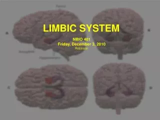
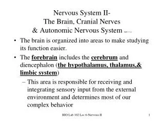
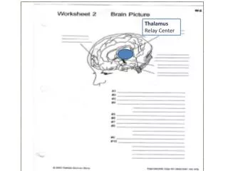
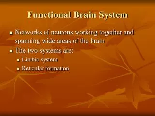
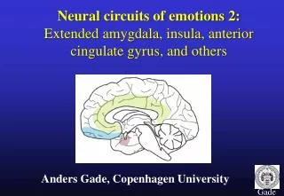
1. The Limbic System: Encircle the upper part of the brain stem.
Is the main �emotional� or �feelings� part of the brain.
Links sensory input to emotions (ex. Recognizing angry facial expressions or bad smells followed by a physical response).
2. Protection of the Brain: Bone-
Done by the skull and the vertebrae.
Meninges-
Provides four functions:
1. Covers and protects the CNS.
2. Protects blood vessels.
3. Contains cerebralspinal fluid.
4. Form partitions in the skull.
3. There are three types of meninges: 1. Dura Mater-
Is the strongest of the meninges.
Made of two layers of fibrous connective tissues.
It is the most superficial layer.
Only found in the skull.
Limits excessive movement of the brain.
2. Arachnoid Mater-
Made of fine elastic connective tissue.
Is the middle layer of the meninges.
Subarachnoid space contains cerebralspinal fluid and large blood vessels.
Provides minimal protection.
3. Pia Mater-
Made of delicate connective tissue.
Full of tiny blood vessels.
Clings tightly to the brain.
Full of tiny blood vessels.
Clings tightly to the brain.
4. Cerebralspinal Fluid: Found in and around brain and spinal cord.
Forms a liquid cushion for CNS.
Helps nourish the brain.
May carry chemical signals through CNS.
Similar in composition as blood plasma.
Volume is ~150ml and is replaced about every 8 hours.
CSF is filtered from blood from the choroid plexures of each of the CNS ventricles.
5. Blood-Brain Barrier: Capillaries of the CNS are selectively permeable to prevent variations in hormones, ions, amino acids, etc.
The hypothalamus has a reduced blood-brain barrier so that it can sample and react to the activity going on in the body.
6. The Spinal Cord: Starts at the base of the skull and extends down to the first or second lumbar vertebra.
Provides a two-way conduction pathway.
Protected by bone, meninges, and cerebralspinal fluid.
Has 31 pairs of spinal nerves.
Important terms associated with the spinal cord:
1. Conus Medullaris- inferior cone-shaped end of the spinal cord.
2. Cervical & Lumbar Enlargements- areas that supply nerves to limbs.
3. Filum Terminale- extension of the pia mater that anchors the inferior end of the spinal cord to the coccyx.
7. Cross-Section Anatomy of the Spinal Cord: Gray matter is found in the core of the cord.
Shaped like the letter �H�
Posterior Horn (sensory)- receives impulses from sensory receptors from dorsal roots and dorsal root ganglia.
Anterior Horn (motor)- sends axons out to effector cells via ventral roots.
Lateral Horn- found in thoracic and lumbar areas of cord. Sends axons out to visceral organs (autonomic).
Gray Commissure- connects the two sides of the horn.
8. Cross-Section Anatomy of the Spinal Cord (cont.): Dorsal and Ventral Roots fuse to form spinal nerves.
Gray matter of spinal cord can be divided into 4 zones:
1. Somatic Sensory
2. Visceral Sensory
3. Somatic Motor
4. Visceral Motor
9. The Peripheral Nervous System: Is the link between the CNS and the outside world.
Includes all neural structures outside of the brain and spinal cord:
A. Sensory Receptors- responds to changes in the environment.
Are modified dendritic endings.
B. Nerves- consist of parallel bundles of peripheral axons enclosed by connective tissue.
10. Nerves (cont.)-
Axons are surrounded by endoneurium (loose connective tissue).
Bundles of these (fascicles) are wrapped with the perineurium (coarser connective tissue).
All the fascicles are enclosed by the epineurium (a tough fibrous sheath).
* Myelin, connective tissue, blood vessels, and lymphatic vessels are also part of the nerve fibers.
There are 3 types of nerves:
Sensory- carry impulses to the CNS.
Motor- carry impulses away from the CNS.
Mixed- contained both types (most common types)