Thrombotic Microangiopathies
850 likes | 1.2k Vues
Explore the historical timeline of Thrombotic Microangiopathies, including key discoveries and features such as vessel wall thickening, endothelial cell detachment, and platelet thrombosis. Learn about the pathogenesis and molecular biology.

Thrombotic Microangiopathies
E N D
Presentation Transcript
Thrombotic Microangiopathies T. Matthew Eison, MD LeBonheur Children’s Hospital Pediatric Nephrology
TMA Timeline -1924: Moschcowitz presented first published example of idiopathic TTP before New York Pathological Society [1,2] -1952: Thrombotic Microangiopathy first described in by Symmers [3] -1965: Familial Atypical HUS first described in combination of hemolytic anemia & azotemia in concordant monozygous twins [4] -1974: Reduced serum levels of complement fraction C3 with normal levels of C4 reported in patients with atypical HUS[5-7] -1976: Bukowski et al published experience w/whole blood exchange transfusion, w/8 /14 TTP patients w/TTP achieving prompt remissions lasting several months to >13 yrs[8] -1981: investigators described two brothers with atypical HUS w/complete complement factor H (CFH) deficiency [9]; parents, who were first cousins, had half-normal CFH levels, indicating inherited defect. -1982: Moake linked von Willebrand factor (VWF) to the pathogenesis of TTP, recognizing circulating “ultralarge” VWF multimers in relapsing acquired or congenital TTP patients in remission [10] -1996: candidate VWF-cleaving protease requiring fluid shear stress or low protein denaturant concentration identified in human plasma by Tsai [11] & independently by Furlan [12] -1997: VWF-cleaving protease demonstrated missing from congenital TTP patients' plasma [13] -1998: Goodship ‘s investigative group [14] showed association b/w atypical HUS & chromosome 1q32 locus, containing genes for CFH and other complement regulators -1998: Adults with acquired idiopathic TTP reported to have severe VWF-cleaving protease deficiency caused by IgG autoantibodies that inhibit the enzyme [15,16] -2001: VWF-cleaving protease purified, cloned, and named ADAMTS13 [17-22] because it belonged to the recently discovered “a disintegrin-like and metalloproteasemetalloprotease with thrombospondin repeats” family of metalloproteases [24] -2001: Mutations in the ADAMTS13 gene shown to cause congenital TTP [21] 12. Furlan M, Robles R, La¨mmle B. Blood.1996;87:4223-4234. 13. Furlan M, Robles R et al. Blood. 1997;89:3097-3103. 14. Warwicker P, Goodship TH, Donne RL, et al. Kidney Int 1998;53:836-44. 15. Tsai HM, Lian EC. N Engl J Med. 1998; 339:1585-1594. 16. Furlan M, Robles R, Galbusera M, et al. N Engl J Med. 1998;339:1578- 1584. 17. Fujikawa K, Suzuki H, McMullen B, Chung D. Blood. 2001;98: 1662-1666. 18. Gerritsen HE, Robles R, Lammle B, Furlan M. Blood. 2001;98:1654-1661. 19. Soejima K, Mimura N, Hirashima M, et al. J Biochem. 2001;130:475-480. 20. Zheng X et al. J Biol Chem. 2001;276: 41059-41063. 21. Levy GG, Nichols WC, Lian EC, et al. Nature. 2001;413:488-494. 22. Hurskainen TL, Hirohata S, Seldin MF, Apte SS. J Biol Chem. 1999;274:25555-25563. 1. Moschcowitz E. 1924;24:21-24. 2. Moschcowitz E. Arch Intern Med. 1925;36:89-93 3. Symmers WStC: Br Med J 2:897–903, 1952 4. Campbell S, Carré IJ. Arch Dis Child 1965;40:654-8. 5. Carreras L, Romero R, Requesens C, et al. JAMA 1981;245:602-4. 6. Stühlinger W, Kourilsky O, Kanfer A, Sraer JD. Lancet 1974;2:788-9. 7. Noris M, Ruggenenti P, Perna A, et al. J Am Soc Nephrol 1999;10:281-93. 8. Bukowski RM, Hewlett JS, Harris JW, et al. Semin Hematol. 1976;13:219-232. 9. Thompson RA, Winterborn MH. Clin Exp Immunol 1981;46:110-9. 10. Moake JL, Rudy CK, Troll JH, et al. N Engl J Med. 1982;307: 1432-1435. 11. Tsai H-M. Blood. 1996;87:4235-4244.
Noris M, Remuzzi G. Hemolytic uremic syndrome. J Am Soc Nephrol. 2005 Apr;16(4):1035-50.
Thrombotic Microangiopathy -pathological term used to describe occlusive microvascular thrombus formation1 -first described in 1952 by Symmers2 -Pathological features: --vessel wall thickening --swelling and detachment of the endothelial cell from the basement membrane --accumulation of material in the subendothelial space --intraluminal platelet thrombosis --partial or complete vessel luminal obstruction and fragmentation of red blood cells 3–6 1. Zheng XL, Sadler JE (2008) Pathogenesis of thrombotic microangiopathies. Annu Rev Pathol 3:249–277 2. Symmers WStC: Thrombotic microangiopathic haemolytic anemia (thrombotic microangiopathy). Br Med J 2:897–903, 1952 3. Ruggenenti P, Noris M, Remuzzi G (2001) Thrombotic microangiopathy, hemolytic uremic syndrome, and thrombotic thrombocytopenic purpura. Kidney Int 60:831–846 4. Copelovitch L, Kaplan BS (2008) The thrombotic microangiopathies. Pediatr Nephrol 23:1761–1767 5. Tsai HM (2006) The molecular biology of thrombotic microangiopathy. Kidney Int 70:16–23 6. Benz K, Amann K (2009) Pathological aspects of membranoproliferative glomerulonephritis (MPGN) and haemolytic uraemic syndrome (HUS)/thrombocytic thrombopenic purpura (TTP).Thromb Haemost 101:265–270
Keir L, Coward RJ. Advances in our understanding of the pathogenesis of glomerular thrombotic microangiopathy. Pediatr Nephrol. 2011 Apr;26(4):523-33
RBCs and fibrin fill up capillary loops in glomerulus in thrombotic microangiopathy (Jones’ silver stain) –Agnes Fogo
Fibrin thrombi extending from glomerulus into arteriole in thrombotic microangiopathy (Jones’ silver stain) –Agnes Fogo
Fibrin tactoids in subendothelial area by EM in thrombotic microangiopathy. –Agnes Fogo
Thrombotic Microangiopathy:Clinical Asociations --consumptive thrombocytopenia --microangiopathic hemolytic anemia --features of organ ischemia1 --symptoms produced depend on the vascular bed and organ affected --most commonly associated with Hemolytic Uremic Syndrome (HUS) and Thrombotic Thrombocytopaenic Purpura (TTP)2 1. Tsai HM (2006) The molecular biology of thrombotic microangiopathy. Kidney Int 70:16–23 2. Zheng XL, Sadler JE (2008) Pathogenesis of thrombotic microangiopathies. Annu Rev Pathol 3:249–277
Is it TTP or HUS? --Hemolytic uremic syndrome (HUS): TMA predominantly seen in the glomeruli; results in acute renal insufficiency* --Thrombotic Thrombocytopenic Purpura (TTP): neurological endothelial cells predominantly affected; therefore neurological features seen* --TTP and HUS often considered together: ----similar etiology ----overlap in clinical symptoms (neurological features in HUS; renal problems in TTP) ----many patients best described as TTP-HUS (Up To Date): severe neurologic sx (seizures & coma) with acute renal failure; *Tsai HM (2006) The molecular biology of thrombotic microangiopathy. Kidney Int 70:16–23
Alternate TTP vs HUS Classification George JN. The thrombotic thrombocytopenic purpura and hemolytic uremic syndromes: overview of pathogenesis (Experience of The Oklahoma TTP-HUS Registry, 1989-2007). Kidney Int Suppl. 2009 Feb;(112):S8-S10.
Besbas N et al; European Paediatric Research Group for HUS. A classification of hemolytic uremic syndrome and thrombotic thrombocytopenic purpura and related disorders. Kidney Int. 2006 Aug;70(3):423-31
Keir L, Coward RJ. Advances in our understanding of the pathogenesis of glomerular thrombotic microangiopathy. Pediatr Nephrol. 2011 Apr;26(4):523-33
Ruggenenti P, Noris M, Remuzzi G (2001) Thrombotic microangiopathy, hemolytic uremic syndrome, and thrombotic thrombocytopenic purpura. Kidney Int 60:831-46.
Thrombotic Thrombocytopenic Purpura:A New Disease Discovery -In Jan.1 & Feb. 19242Moschcowitzpresented 1st known case before New York Pathological Society: “a hitherto undescribed disease” “remarkable, clinically and anatomically” -Case: Healthy 16-year-old girl w/initial sx ----sudden arm weaknessarms ----pain w/wrist and elbow movement ----pallor ----fever (38°C-39°C) -Symptoms worsened; Day #10 illness, admitted w/findings ----anemia ----leukocytosis ----a few petechiae on one arm ----occult blood in gastric contents and stool ----nl serum creatinine -Day #14 illness: ----mild left hemiparesis and facial paralysis -Day #15 illness: ----became comatose and died -On limited autopsy: hyaline thrombi in terminal arterioles and capillaries of heart, kidney, spleen, and liver; the lungs were spared -No platelet count obtained; -no report of schistocytes; -therefore not complete description of thrombocytopenia or microangiopathic hemolytic anemia (Nonetheless, recognized as first published example of idiopathic TTP) 1. Moschcowitz E. Proc N Y Pathol Soc. 1924;24:21-24. 2. Moschcowitz E. Arch Intern Med. 1925;36:89-93. 3. Sadler JE. Blood. 2008 Jul 1;112(1):11-8.
Thrombotic Thrombocytopenic Purpura (TTP):Epidemiology -more common in females age 10-39 yrs -highest incidence is seen in fourth decade -annual incidence 3.7 cases per 1,000,0001 1. Michael M, Elliott EJ, Ridley GF, Hodson EM, Craig JC (2009) Interventions for haemolytic uraemic syndrome and thrombotic thrombocytopenic purpura. Cochrane Database Syst Rev CD003595 2. Keir L, Coward RJ. Advances in our understanding of the pathogenesis of glomerular thrombotic microangiopathy. Pediatr Nephrol. 2011 Apr;26(4):523-33
Thrombotic Thrombocytopenic Purpura (TTP):Clinical Presentation -Classic "Pentad" of symptoms: ----microangiopathic hemolytic anemia ----thrombocytopaenia ----neurological symptoms ----renal damage ----fever -Previously a diagnosis of exclusion; now ADAMTS13 activity assay in use1 -Plasma exchange improves symptoms; now standard treatment for TTP2 1. Sadler JE (2008) Von Willebrand factor, ADAMTS13, and thrombotic thrombocytopenic purpura. Blood 112:11–18 2. Zheng XL, Sadler JE (2008) Pathogenesis of thrombotic microangiopathies. Annu Rev Pathol 3:249–277 3. Keir L, Coward RJ. Advances in our understanding of the pathogenesis of glomerular thrombotic microangiopathy. Pediatr Nephrol. 2011 Apr;26(4):523-33
George JN. The thrombotic thrombocytopenic purpura and hemolytic uremic syndromes: overview of pathogenesis (Experience of The Oklahoma TTP-HUS Registry, 1989-2007). Kidney Int Suppl. 2009 Feb;(112):S8-S10.
Vesely SK, George JN, Lämmle B, Studt JD, Alberio L, El-Harake MA, Raskob GE. ADAMTS13 activity in thrombotic thrombocytopenic purpura-hemolytic uremic syndrome: relation to presenting features and clinical outcomes in a prospective cohort of 142 patients. Blood. 2003 Jul 1;102(1):60-8
George JN. The thrombotic thrombocytopenic purpura and hemolytic uremic syndromes: overview of pathogenesis (Experience of The Oklahoma TTP-HUS Registry, 1989-2007). Kidney Int Suppl. 2009 Feb;(112):S8-S10.
Complications of Plasma Exchange in TTP In a study of 57 consecutive patients from 2002-2005 from the Oklahoma TTP-HUS Registry: Howard MA, Williams LA, Terrell DR, Duvall D, Vesely SK, George JN. Complications of plasma exchange in patients treated for clinically suspected thrombotic thrombocytopenic purpura-hemolytic uremic syndrome. Transfusion. 2006 Jan;46(1):154-6.
Complications of Plasma Exchange in TTP Howard MA, Williams LA, Terrell DR, Duvall D, Vesely SK, George JN. Complications of plasma exchange in patients treated for clinically suspected thrombotic thrombocytopenic purpura-hemolytic uremic syndrome. Transfusion. 2006 Jan;46(1):154-6.
Thrombotic Thrombocytopenic Purpura (TTP):History of Pathogenesis Elucidation -TTP treated successfully x 20 yrs w/plasma exchange before mechanism understood1 -In 1982, TTP patients recognized to have circulating ultra-large multimers of von Willebrand factor during remission (as multimers not in healthy people, TTP patients were hypothesized to lack protease to cleave ultra-large multimers)2 -TTP now known to be disorder of von Willebrand factor (vWF) regulation3 1. Moake J (2009) Thrombotic thrombocytopenia purpura (TTP) and other thrombotic microangiopathies. Best Pract Res Clin Haematol 22:567–576 2. Sadler JE (2008) Von Willebrand factor, ADAMTS13, and thrombotic thrombocytopenic purpura. Blood 112:11–18 3. Tsai HM (2006) The molecular biology of thrombotic microangiopathy. Kidney Int 70:16–23 4. Keir L, Coward RJ. Advances in our understanding of the pathogenesis of glomerular thrombotic microangiopathy. Pediatr Nephrol. 2011 Apr;26(4):523-33
Thrombotic Thrombocytopenic Purpura (TTP) Pathogenesis: von Willebrand factor (vWF) -glycoprotein produced by endothelial cells -regulates platelet aggregation and adhesion -released from endothelial cells as ultralarge multimers (UL-vWF) when vascular injury occurs -Some multimers remain associated with endothelial cell surface (providing platelet-binding sites) -Multimers may bind other blood components too, (e.g. leukocytes) -Platelet binding to UL-vWF regulated by the metalloprotease ADAMTS13 (a-disintegrin-like and metalloprotease and thrombospondin repeats) -deficiency of ADAMTS 13 accounts for majority of patients with congenital TTP -majority of acquired cases occur due to antibody formation against ADAMTS 131 1. Sadler JE (2008) Von Willebrand factor, ADAMTS13, and thrombotic thrombocytopenic purpura. Blood 112:11–18 2. Keir L, Coward RJ. Advances in our understanding of the pathogenesis of glomerular thrombotic microangiopathy. Pediatr Nephrol. 2011 Apr;26(4):523-33
Sadler JE. Von Willebrand factor, ADAMTS13, and thrombotic thrombocytopenic purpura. Blood. 2008 Jul 1;112(1):11-8.
Sadler JE. Von Willebrand factor, ADAMTS13, and thrombotic thrombocytopenic purpura. Blood. 2008 Jul 1;112(1):11-8.
Vesely SK, George JN, Lämmle B, Studt JD, Alberio L, El-Harake MA, Raskob GE. ADAMTS13 activity in thrombotic thrombocytopenic purpura-hemolytic uremic syndrome: relation to presenting features and clinical outcomes in a prospective cohort of 142 patients. Blood. 2003 Jul 1;102(1):60-8
Hemolytic uremic syndrome (HUS):Epidemiology -More commonly seen in children rather than adults -most common cause of pediatric acute renal failure -occurs most frequently in children under the age of 5 years -annual incidence of 6.1 cases per 100,000 children under 5 years -affects 0.2 - 4.28 people per 100,000 worldwide1 1. Serna A 4th, Boedeker EC (2008) Pathogenesis and treatment of Shiga toxin-producing Escherichia coli infections. Curr Opin Gastroenterol 24:38–47 2. Noris M, Remuzzi 1. G. Hemolytic uremic syndrome. J Am Soc Nephrol 2005; 16:1035-50
Hemolytic Uremic Syndrome Classification -A) Typical -More than 90% of cases in children -secondary to infection with ---1) Escherichia coli serotypes O157:H7, O111:H8, O103:H2, O123, O26, or others, which produce Shiga-like toxin (Stx) 1 ---2) several other bacteria, such as Streptococcus pneumoniae.2 -B) Atypical -Approximately 10% of cases in children -not caused by either Stx-producing bacteria or streptococci.1,3 -poor prognosis: ---1) death rates as high as 25%3 ---2) progression to end-stage renal disease in half the patients.1,2 -Research has linked to uncontrolled activation of the complement system. -more commonly associated with extra-renal effects than typical HUS4 *Stx-related lesions indistinguishable from atypical form via standard histologic analysis. 4,5 1. Noris M, Remuzzi 1. J Am Soc Nephrol 2005; 16:1035-50. 2. Constantinescu AR, Bitzan M, Weiss LS, et al. Am J Kidney Dis 2004;43:976-82. 3. Kaplan BS, Meyers KE, Schulman SL. J Am Soc Nephrol 1998;9:1126-33. 4. Ruggenenti P, Noris M, Remuzzi G. Kidney Int 2001;60: 831-46. 5. Benz K, Amann K. Thromb Haemost 2009;101:265-70.
Shigatoxin/Diarrhea-associated HUS:Epidemiology -Most common cause of HUS1 -predominantly a disease of childhood, but can also affect adults, particularly the elderly -It is fatal in 3–5% of cases -overall incidence 2.1 cases per 100,000 persons/yr 2 -peak incidence in children younger than 5 yr (6.1 per 100,000/yr)2 -lowest incidence in adults 50 to 59 yr of age (0.5 per 100,000/yr)2 -incidence parallels seasonal fluctuation of E. coli O157:H7 infections [peak in warmer months (June-September)] -In U.S., causes approx. 70,000 illnesses & 60 deaths annually3 -In Argentina & Uruguay, E. coli infections endemic; estimated Stx-HUS incidence 10.5 per 100,000/yr4 1. Scheiring J, Andreoli SP, Zimmerhackl LB (2008) Pediatr Nephrol 23:1749–1760 2. Ruggenenti P, Noris M, Remuzzi G: Kidney Int 60: 831–846, 2001 3. Mead PS, Slutsker L, Dietz V, McCaig LF, Bresee JS, Shapiro C, Griffin PM, Tauxe RV: Emerg Infect Dis 5: 607–625, 1999 4. Meichtri L, Miliwebsky E, Gioffre A, Chinen I, Baschkier A, Chillemi G, Guth BE, Masana MO, Cataldi A, Rodriguez HR, Rivas M: Int J Food Microbiol 96: 189–198, 2004 5. Keir L, Coward RJ. Pediatr Nephrol. 2011 Apr;26(4):523-33 6. Noris M, Remuzzi G. J Am Soc Nephrol. 2005 Apr;16(4):1035-50.
Shigatoxin/Diarrhea-associated HUS:Source of Infection Vectors for Stx-producing E. coli transmission1,2 : ---Cattle (colonize healthy cattle intestine) ---deer ---sheep ---goats ---horses ---dogs ---birds ---flies Sources of infection in humans ---manure and water troughs in farms (therefore increased risk of infection in rural areas) ---contaminated milk ---contaminated meat (contamination occurs at slaughter; bacteria internalized during grinding; survives cooking)3 ---unchlorinated water4 ---contact with infected animals or humans excrement3,5,6 ---environmental contamination7 ---Contaminated fruits and vegetables (radish sprouts, lettuce, apple cider) ---Unpasteurized apple juice in several outbreaks8 ---Person-to-person (child care and long-term care facilities)3 1. Ruggenenti P, Noris M, Remuzzi G: Kidney Int 60: 831–846, 2001 2. Griffin PM, Tauxe RV: Epidemiol Rev 13: 60–98, 1991 3. Mead PS, Griffin PM: Lancet 352: 1207–1212, 1998 4. McCarthy TA et al: Pediatrics 108: E59, 2001 5. Locking ME et al: Epidemiol Infect 127: 215–220, 2001 6. Mead PS et al: Arch Intern Med 157: 204–208, 1997 7. Varma JK et al: JAMA 290: 2709–2712, 2003 8. Cody SH et al: Ann Intern Med 130: 202–209, 1999 9. Noris M, Remuzzi G. J Am Soc Nephrol. 2005 Apr;16(4):1035-50
Shigatoxin/Diarrhea-associated HUS:Clinical Presentation -Avg interval E. coli exposure to illness 3 days (range, 1 to 8)1 -Typical initial presentation of abdominal cramps & nonbloody diarrhea -Diarrhea hemorrhagic in 70% of cases; usually within 1 or 2 days1 -2 to 5 days post diarrhea onset with symptoms of pallor, weakness, oligo-anuria -HUS usually diagnosed 6 days post diarrhea onset2 -Symptom Associations: Vomiting (30-60%); fever (30%), elevated WBC -Barium Enema: “thumb-printing” (edema & submucosal hemorrhage, especially in ascending & transverse colon)2 1. Chandler WL, Jelacic S, Boster DR, Ciol MA, Williams GD, Watkins SL, Igarashi T, Tarr PI: Prothrombotic coagulation abnormalities preceding the hemolytic-uremic syndrome. N Engl J Med 346: 23–32, 2002 2. Ruggenenti P, Noris M, Remuzzi G: Thrombotic microangiopathy, hemolytic uremic syndrome, and thrombotic thrombocytopenic purpura. Kidney Int 60: 831–846, 2001 3. Keir L, Coward RJ. Pediatr Nephrol. 2011 Apr;26(4):523-33 4. Noris M, Remuzzi G. J Am Soc Nephrol. 2005 Apr;16(4):1035-50.
Shigatoxin/Diarrhea-associated HUS:Risk of Developing HUS -Only 10–15% of enterohaemorrhagic E. coli- infected children develop HUS1 -Associated with increased risk of HUS post E. coli infection2: ---diarrhea ---fever ---vomiting ---elevated leukocyte count ---extremes of age ---female gender ---antimotility agents3 1. Keir L, Coward RJ. Pediatr Nephrol. 2011 Apr;26(4):523-33 2. Mead PS, Griffin PM: Escherichia coli O157:H7. Lancet 352: 1207–1212, 1998 3. Beatty ME, Griffin PM, Tulu AN, Olsen SJ: Culturing practices and antibiotic use in children with diarrhea. Pediatrics 113: 628–629, 2004 4. Noris M, Remuzzi G. J Am Soc Nephrol. 2005 Apr;16(4):1035-50
Shigatoxin/Diarrhea-associated HUS:Diagnosis -Stx-E. coli may shed in stool several weeks post symptoms resolution, particularly in children <5 yrs of age1 -Diagnosis depends on stool culture-detection of Stx-E. coli -Research laboratories can perform serologic tests for Stx- and O157 LPS- antibodies -Rapid detection tests of stool for E. coli O157:H7 & Stx being developed 1) Ruggenenti P, Noris M, Remuzzi G: Thrombotic microangiopathy, hemolytic uremic syndrome, and thrombotic thrombocytopenic purpura. Kidney Int 60: 831–846, 2001 2) Noris M, Remuzzi G. J Am Soc Nephrol. 2005 Apr;16(4):1035-50.
Shigatoxin/Diarrhea-associated HUS:Sequelae -50-75% diarrhea-associated HUS (D + HUS) cases require dialysis therapy in the acute phase1 -70% of HUS patients require RBC transfusions -25% have neurologic involvement, including stroke, seizure, and coma2,3,4 -Despite ICU & Dialysis, 3-5% of Stx-HUS patients die in acute phase2 -12% risk of death or permanent ESRD, 25% risk of GFR < 80 ml/min/1.73 m2 (Meta-analysis of 3476 patients from 49 published studies, mean follow-up 4.4 yr)4 -Associated with poor long-term prognosis: severe acute illness, CNS symptoms, need for initial dialysis 4,5 -Mortality rate ~ 30% when associated with S. dysenteriae infection6 (often complicated by bacteremia w/septic shock, DIC, acute cortical necrosis) 1. Scheiring J, Andreoli SP, Zimmerhackl LB (2008) Pediatr Nephrol 23:1749–1760 2. Milford D: The hemolytic uremic syndromes in the United Kingdom. In: Hemolytic Uremic Syndrome and Thrombotic Thrombocytopenic Purpura, edited by Kaplan BS, Trompeter RS, Moake JL, New York, Marcel Dekker, 1992, pp 39–59 3. Mead PS, Griffin PM: Escherichia coli O157:H7. Lancet 352: 1207–1212, 1998 4. Garg AX, et al. JAMA 290: 1360–1370, 2003 5. Tonshoff B, Sammet A, Sanden I, Mehls O, Waldherr R, Scharer K: Nephron 68: 63–70, 1994 6. Date A, Raghupathy P, Jadhav M, Pereira SM, Shastry JC: Ann Trop Paediatr 2: 1–6, 1982 7. Keir L, Coward RJ. Pediatr Nephrol. 2011 Apr;26(4):523-33 8. Noris M, Remuzzi G. J Am Soc Nephrol. 2005 Apr;16(4):1035-50.
Shigatoxin/Diarrhea-associated HUS:Pathogenesis -Stx-producing serotypes (E. coli O157:H7, O111, nonmotile O26:H11, O103:H2) may produce large amts of Stx -Stx traverse polarized gastrointestinal epithelial cells via transcellular pathways1, to systemic circulation (However, Free toxin has never been detected HUS patient's circulation) -Shiga toxin's high-affinity cellular target: Globotriaosylceramide (Gb3)2 -Gb3: glycosphingolipid receptor on human endothelial cells, podocytes and tubular cells -Podocytes, proximal tubule cells, glomerular endothelial cells all sensitive to Stx cytotoxicity. -In vitro, Stx rapidly binds with low-affinity to polymorphonuclear leukocytes (PMNs) (not erythrocytes, monocytes, platelets, or lipoproteins)3 -PMN Stx receptor 100-fold lower affinity than high-affinity receptor Gb3 expressed on glomerular endothelial cells. -in in vitro cocultures, PMNs Stx to glomerular endothelial cells, which promote cell death3 (in vivo would explain rapid clearance of Stx from circulation and release to target organs) 1. AchesonDWK,MooreR,DeBreuler S: Infect Immunol 64:3294–3300, 1996 2. Psotka MA, Obata F, Kolling GL, Gross LK, Saleem MA, Satchell SC, Mathieson PW, Obrig TG (2009) Shiga toxin 2 targets the murine renal collecting duct epithelium. Infect Immun 77:959–969 3. te Loo DMWM, Monnens LAH, van der Velden TJAM, et al: Blood 95:3396–3402, 2000 4. Ruggenenti P, Noris M, Remuzzi G. Kidney Int. 2001 Sep;60(3):831-46. 5. Keir L, Coward RJ. Pediatr Nephrol. 2011 Apr;26(4):523-33
Streptococcal pneumoniae-related HUS:Epidemiology -accounts for 5% of childhood cases of HUS1 -incidence of HUS following pneumococcal infection estimated to be 0.4–0.6% -Children under 2 years are most commonly affected 1. Copelovitch L, Kaplan BS (2008) Streptococcus pneumoniae associated hemolytic uremic syndrome. Pediatr Nephrol 23:1951– 1956 2. Keir L, Coward RJ. Advances in our understanding of the pathogenesis of glomerular thrombotic microangiopathy. Pediatr Nephrol. 2011 Apr;26(4):523-33
Streptococcal pneumoniae-related HUS:Clinical Presentation -follows invasive pneumococcal disease -linked to having a high bacterial load -HUS usually develops 3–13 days post initial infection -associated with a longer period of oligoanuria & acute dialysis period than stx HUS -10% of patients progress to ESRD; 12% mortality -highest mortality related to S.pneumoniae meningitis complicated by HUS Keir L, Coward RJ. Advances in our understanding of the pathogenesis of glomerular thrombotic microangiopathy. Pediatr Nephrol. 2011 Apr;26(4):523-33
Streptococcal pneumoniae-related HUS Pathogenesis: Thomsen–Friedenreich (TF) crypt antigen -found on surface of erythrocytes, platelets and glomerular endothelial cells -normally masked by neuraminic acid -neuraminidase (produced by all serotypes of S. pneumoniae) cleaves n-acetyl neuraminic acid from cell surface, exposing TF antigen -host in response produces IgM antibodies, which bind to the TF antigen -Immune response cascade ensues, culminating in HUS (red cells, platelets, glomerular endothelial cell damaged) -TF antigen also found on hepatocytes (explains hepatic dysfunction in some patients) Keir L, Coward RJ. Advances in our understanding of the pathogenesis of glomerular thrombotic microangiopathy. Pediatr Nephrol. 2011 Apr;26(4):523-33
Atypical Hemolytic Uremic Syndrome:Classification A) Familial Form -less than 20% of cases -poor prognosis: end-stage renal disease or death of 50 to 80% -First described in 1965: combination of hemolytic anemia & azotemia in concordant monozygous twins.1 -Typically reported in children but infrequently, in adults -Both autosomal dominant and recessive patterns of inheritance. 2 1. Campbell S, Carré IJ. Fatal haemolytic uraemic syndrome and idiopathic hyperlipaemia in monozygotic twins. Arch Dis Child 1965;40:654-8. 2. Kaplan BS, Chesney RW, Drummond KN. Hemolytic uremic syndrome in families.N Engl J Med 1975;292:1090-3.
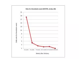
Atypical Hemolytic Uremic Syndrome:Classification(cont’d) B) Sporadic Form -Designation when no family history of the disease -50% of cases appear to be idiopathic. -Triggers: --1) human immunodeficiency virus --2) cancer --3) organ transplantation ----de novo TMA reported in 3.6 to 14.0% of all kidney transplant recipients; associated with humoral rejection & calcineurin inhibitors.1-3 --4) pregnancy ---- ~10 to 15% of female patients with atypical HUS develops during pregnancy or post partum.4,5 --5) Medications: ----a) anticancer drugs ----b) immunotherapeutic agents (e.g., cyclosporine and tacrolimus) ----c) antiplatelet agents (e.g., ticlopidine and clopidogrel). 1. Ruggenenti P. Kidney Int 2002; 62:1093-104. 2. Karthikeyan V, Parasuraman R, Shah V, Vera E, Venkat KK. Am J Transplant 2003;3:1289-94. 3. Zarifian A, Meleg-Smith S, O’Donovan R, Tesi RJ, Batuman V. Kidney Int 1999;55:2457-66. 4. Noris M, Remuzzi 1. G. Hemolytic uremic syndrome. J Am Soc Nephrol 2005; 16:1035-50. 5. Besbas N, Karpman D, Landau D, et al. Kidney Int 2006;70:423-31. 6. Ruggenenti P, Noris M, Remuzzi G. Kidney Int 2001;60: 831-46. 7. Zakarija A, Bennett C. Semin Thromb Hemost 2005;31:681-90.
Atypical HUS Association with Complement System Abnormalities -Since 1974, reduced serum levels of complement fraction C3 with normal levels of C4 reported in patients with atypical HUS, reflecting complement activation and consumption.1-3 -Genetic abnormalities in complement system proteins documented in both familial and sporadic forms 4-11 -Two patients with Stx-related HUS reported with mutations in complement regulatory genes 10,12 -Mutations in complement pathway have been found to account for 50 to 60% of cases of atypical HUS13 1. Carreras L, Romero R, Requesens C, et al. JAMA 1981;245:602-4. 2. Stühlinger W, Kourilsky O, Kanfer A, Sraer JD. Lancet 1974;2:788-9. 3. Noris M, Ruggenenti P, Perna A, et al. J Am Soc Nephrol 1999;10:281-93. 4. Caprioli J, Noris M, Brioschi S, et al. Blood 2006;108:1267-79. 5. Noris M, Brioschi S, Caprioli J, et al. Lancet 2003;362:1542-7. 6. Richards A, Kemp EJ, Liszewski MK, et al. Proc Natl Acad Sci U S A 2003;100:12966-71. 7. Goicoechea de Jorge E, Harris CL, Esparza-Gordillo J, et al. Proc Natl Acad Sci U S A 2007;104:240-5. 8. Frémeaux-Bacchi V, Miller EC, Liszewski MK, et al. Blood 2008; 112:4948-52. 9. Le Quintrec M, Lionet A, Kamar N, et al. Am J Transplant 2008;8:1694-701. 10. Fang CJ, Fremeaux-Bacchi V, Liszewski MK, et al. Blood 2008;111:624-32. 11. Fakhouri F, Jablonski M, Lepercq J, et al. Blood 2008;112: 4542-5. 12. Loirat C, Noris M, Fremeaux-Bacchi V. Pediatr Nephrol 2008;23:1957-72. 13. Noris M, Remuzzi G. N Engl J Med. 2009;361:1676-87
Overview of Complement System in Hemolytic Uremic Syndrome -Consists of plasma and membrane-bound proteins that protect against invading organisms.1 -Composed of three activation pathways — classic, lectin, and alternative -Pathways produce protease complexes termed C3 and C5 convertases that cleave C3 and C5, leading to the membrane-attack complex -C3 convertases of the classic and lectin pathways are formed by C2 and C4 fragments -Alternative pathway convertase cleaves C3 but not C4.1 -C3 hydrolysis in plasma initiates the alternative pathway, leading to the deposition of C3b onto almost all plasma-exposed surfaces 2 1. Walport MJ. Complement: first of two parts. N Engl J Med 2001;344:1058-66. 2. Devaux P, Christiansen D, Fontaine M, Gerlier D. Control of C3b and C5b deposition by CD46 (membrane cofactor protein) after alternative but not classical complement activation. Eur J Immunol 1999; 29:815-22.
Noris M, Remuzzi G. Hemolytic uremic syndrome. J Am Soc Nephrol. 2005 Apr;16(4):1035-50.
Overview of Complement System in Hemolytic Uremic Syndrome (cont’d) -On host cells, membrane-anchored and fluid-phase regulators control complement activation : ----favoring the cleavage of C3b to inactive C3b (iC3b) by complement factor I (CFI) activity ----dissociating the multicomponent C3 and C5 convertases (i.e., decay-acceleration activity). -Without normal regulation, C3b deposition: ----increases by more than a factor of 20 through the amplification loop1 ----causes activation of the complement cascade, which remains active until complement components are consumed. -Foreign targets and injured cells are attacked by complement due to: ----a) inability to bind soluble regulators ----b) no membrane-bound regulators . -On the surface of bacteria, C3b binds to specific receptors on neutrophils and macrophages, resulting in phagocytosis of complement-tagged bacteria. -Therefore in a patient with atypical HUS, low serum C3 level with normal C4 level indicates selective alternative pathway activation 2 1. Devaux P, Christiansen D, Fontaine M, Gerlier D. Control of C3b and C5b deposition by CD46 (membrane cofactor protein) after alternative but not classical complement activation. Eur J Immunol 1999; 29:815-22. 2. Noris M, Ruggenenti P, Perna A, et al. Hypocomplementemia discloses genetic predisposition to hemolytic uremic syndrome and thrombotic thrombocytopenic purpura: role of factor H abnormalities. J Am Soc Nephrol 1999;10:281-93.
Keir L, Coward RJ. Advances in our understanding of the pathogenesis of glomerular thrombotic microangiopathy. Pediatr Nephrol. 2011 Apr;26(4):523-33
Keir L, Coward RJ. Advances in our understanding of the pathogenesis of glomerular thrombotic microangiopathy. Pediatr Nephrol. 2011 Apr;26(4):523-33
Laboratory & Pathological Evidence of Complement Abnormalities in Atypical HUS -HUS patients w/low serum C3 levels have high levels of activated complement components, including C3b, C3c, and C3d.1 -Granular C3 deposits in glomeruli & arterioles during acute disease C/W activation of complement & local C3 consumption. 2,3 -C9 staining in glomeruli & small arteries with intimal proliferation and thrombosis suggests activation up to the final lytic C5b-9 membrane-attack complex.4 1. Kim Y, Miller K, Michael AF. Breakdown products of C3 and factor B in hemolytic-uremic syndrome. J Lab Clin Med 1977;89:845-50. 2. Stühlinger W, Kourilsky O, Kanfer A, Sraer JD. Haemolytic-uraemic syndrome: evidence for intravascular C3 activation. Lancet 1974;2:788-9. 3. Barré P, Kaplan BS, de Chadarévian JP, Drummond KN. Hemolytic uremic syndrome with hypocomplementemia, serum C3NeF, and glomerular deposits of C3. Arch Pathol Lab Med 1977;101:357-61. 4. Landau D, Shalev H, Levy-Finer G, Polonsky A, Segev Y, Katchko L. Familial hemolytic uremic syndrome associated with complement factor H deficiency. J Pediatr 2001;138:412-7.