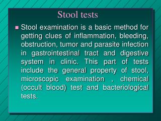
Stool Analysis
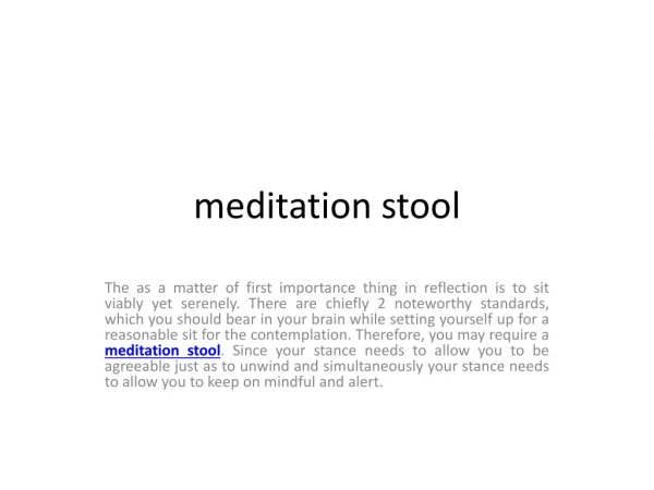
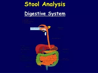
Stool Analysis. A stool analysis is a series of tests done on a stool (feces) sample to help diagnose certain conditions affecting the digestive tract . These conditions can include infection (such as from parasites , viruses , or bacteria ), poor nutrient absorption, or cancer.

Stool Analysis
E N D
Presentation Transcript
Stool Analysis A stool analysis is a series of tests done on a stool (feces) sample to help diagnose certain conditions affecting the digestive tract. These conditions can include infection (such as from parasites, viruses, or bacteria), poor nutrient absorption, or cancer. General view of digestive system: The digestive tract is a hollow tube that extends through the center of the body from the mouth to the anus
Digestive System The digestive system consist of two parts: the Alimentary tract (Gastro Intestinal Tract) (GIT) and the accessory glands .Fig (1) Functions: process food that is placed in the mouth and that passes through the digestive tract where it is broken down, processed, absorbed and utilized as energy that fuels the body's daily functions . The excess of dietary mass and any toxins or waste products that are produced through metabolic processes are excreted through the anus. Organs that are indirectly involved are known as the accessory digestive organs.
Anatomyof the Digestive System • The digestive system consists of two main parts: • 1-The gastrointestinal tract (GIT) • This tract is divided into the upper and lower gastrointestinal tracts: • Upper Gastrointestinal Tract • The upper gastrointestinal tract includes: • Oral cavity(Mouth) • Pharynx • Esophagus: the fibro muscular tube through which food passes, aided by peristaltic contractions, from the pharynx to the stomach. • Stomach: secretes protein-digesting enzymes called proteases and strong acids to aid in food digestion, before sending partially digested food to the small intestines. • Duodenum: the first section of the small intestine and may be the principal site for iron absorption • Lower Gastrointestinal Tract • The lower gastrointestinal tract includes most of the small intestine and all of the large intestine. According to some sources, it also includes the anus.
-The small intestine has three parts: • Duodenum:Here the digestive juices from the pancreas (digestive enzymes) and the gallbladder (bile) mix together. The digestive enzymes break down proteins and bile emulsify fats into micelles. The duodenum contains Brunner's glands which produce bicarbonate, and pancreatic juice contains bicarbonate to neutralize hydrochloric acid of the stomach. • Jejunum: This is the midsection of the intestine, connecting the duodenum to the ileum. It contains the plicae circulars and villi to increase the surface area of that part of the GI Tract. • Ileum:Has villi, where all soluble molecules are absorbed into the blood (capillaries and lacteals). • -The large intestine has four parts: • Cecum:The vermiform appendix is attached to the cecum. • Colon: Includes the ascending colon, transverse colon, descending colon, and sigmoid flexure. The main function of the colon is to absorb water, but it also contains bacteria that produce beneficial vitamins like vitamin K. • Rectum • Anus • 2-Accessory glands • The accessory glands consist of thesalivarygland, liver, pancreas and gall bladder
Function of digestive system • The digestive system responsible for consuming and digesting foodstuffs, absorbing nutrients, and expelling waste.
Immune barrier • The gastrointestinal tract is also a prominent part of the immune system. The surface area of the digestive tract is estimated to be the surface area of a football field. With such a large exposure, the immune system must work hard to prevent pathogens from entering into blood and lymph. • The low pH(ranging from 1 to 4) of the stomach is fatal for many microorganisms that enter it. Similarly, mucus (containing IgAantibodies) neutralizes many of these microorganisms. Other factors in the GI tract help with immune function as well, including enzymes in saliva and bile • Health-enhancing intestinal bacteria of the gut flora serve to prevent the overgrowth of potentially harmful bacteria in the gut. These two types of bacteria compete for space and "food," as there is limited resources within the intestinal tract. A ratio of 80-85% beneficial to 15-20% potentially harmful bacteria generally is considered normal within the intestines.
Normal Gastrointestinal Microbiota (normal flora) • Bacteria make up most of the flora in the colon and up to 60% of the dry mass of feces. Somewhere between 300 and 1000 different species live in the gut. Fungi and protozoa also make up a part of the gut flora, but little is known about their activities. • The microorganisms perform a host of useful functions, such as: • fermenting unused energy substrates, training the immune system, preventing growth of harmful, pathogenic bacteria, regulating the development of the gut, producing vitamins for the host (such as biotin and vitamin K), and producing hormones to direct the host to store fats.
Over 99% of the bacteria in the gut are anaerobes, but in the cecum, aerobic bacteria reach high densities. Not all the species in the gut have been identified because most cannot be cultured, and identification is difficult. Populations of species vary widely among different individuals but stay fairly constant within an individual over time, even though some alterations may occur with changes in lifestyle, diet and age. • Most bacteria belong to the genera Bacteroides, Clostridium, Fusobacterium, Eubacterium, Ruminococcus, Peptococcus, Peptostreptococcus, and Bifidobacterium. Other genera, such as Escherichia and Lactobacillus, are present to a lesser extent. Species from the genus Bacteroides alone constitute about 30% of all bacteria in the gut, suggesting that this genus is especially important in the functioning of the host. The currently known genera of fungi of the gut flora include Candida, Saccharomyces, Aspergillus, and Penicillium.
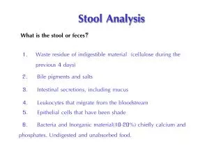
Stool or Feces Define as: Awastes of food digestion which did not absorbed. Normal stool characteristics: Brown in colour, soft, homogenized, do not contain blood, mucus, pus, bacteria, fungi, parasites or viruses, looks like cylinder , PH=6, contain less than 25mg/g stool reducing factors and its quantity around 200g/day.
Diseases of digestive system • There are a number of diseases and conditions affecting the gastrointestinal system, including: • Infection:Gastroenteritis is an inflammation of the intestines. It occurs more frequently than any other disease of the intestines. • -Classification of infection in the digestive system • Infections in the digestive system are classified in two groups: • Exogenous infections –pathogens that come into the body • Endogenous infections –organisms that are part of the normal microbial flora
Exogenous infections • Exogenous infections are brought in with contaminated food or water. • C. difficile and other exogenous infections are frequently acquired in hospital environments. • Helicobacter pylori spreads through oral-oral or fecal-oral contact. • Exogenous infections can cause nausea and vomiting within 6 hours. • Endogenous infections • Endogenous infections are caused by organisms that are part of the normal flora. • Streptococcus andEnterococcus are examples.
Cancer: may occur at any point in the gastrointestinal tract, and includes mouth cancer, tongue cancer, esophageal cancer, stomach cancer, and colorectal cancer. • Inflammatory conditions: Ileitis is an inflammation of the ileum; Colitis is an inflammation of the intestine, Appendicitis:Is inflammation of the vermiform appendix located at the caecum. This is a potentially fatal condition if left untreated; most cases of appendicitis require surgical intervention. • Diverticular disease:Is a condition that is very common in older people. It usually affects the large intestine and the small intestine as well. Diverticulosis occurs when pouches form on the intestinal wall. Once the pouches become inflamed it is known as diverticulitis, (the patients stool is red and bloody). • Cholelithiasis, gallstones in the gallbladder • Peptic ulcer, open sore in the lining of the stomach or duodenum • Anal fistula, abnormal tube-like passageway near the anus. • Dysentery, painful, inflamed intestines commonly caused by bacterial infection.
Hemorrhoids, swollen, twisted, varicose veins in the rectal region
Symptoms • Several symptoms are used to indicate problems with the gastrointestinal tract: • Nausea, unpleasant sensation in the stomach associated with a tendency to vomiting. • Vomiting, which may include regurgitation of food (due to GIT inflammation, acute pain, drugs, pregnancy, emotions)or the vomiting of blood (as in upper GIT bleeding (Haematomesis)). • Melena, black, tarry stools; feces containing digested blood (which isa sign of upper GIT bleeding). • Diarrhoea,the passage of liquid or more frequent stools (watery), more than three times in a day and more than 200g/day, and with incontinence. There are two types of diarrhea: Acute and chronic diarrhea. • -Acute Diarrhoea : short in time , do not need any medication unless the patient is immunecomprised. Most cases (90%) are due to ingestion of contaminated food with bacteria or its toxin ,viruses and parasites, the rest (10%) are due to medication drugs like antibiotics. • -Chronic Diarrhoea:long in time (more than one month), need medication for dehydration because of losing K, Mg, Na salts which may cause death. The causes of chronic Diarrhoea are colitis, malabsorption, colon cancer, irritation of small intestine with some drugs.
Constipation,which refers to the passage of fewer and hardened stools (difficulty in passing stools (feces)), due to: pregnancy, GITobstructions with tumors, diverticulum and hemorrhoids, Age. • Dysphagia, difficulty in swallowing • Eructation, gas expelled from the stomach through the mouth • Flatus, gas expelled through the anus. • Jaundice (icterus), yellow-orange coloration of the skin and whites of the eyes caused by high levels of bilirubin in the blood (hyperbilirubinemia) • Statorrhea, fat in the feces; frothy, foul-smelling fecal matter, due to malabsorption which result from pancreatic diseases.
Laboratory examination of feces (General Stool Examination) (GST) • Collection of the stool specimen: • To ensure that good specimens are provided for examination, it is important to note the following: • 1. A clean dry container must be used for the collection of fecal samples. Urine and water will destroy trophoziot, if present, and the presence of dirt also causes identification problems. • 2. Ideally the specimen should be brought to the lab as soon as it is passed, to avoid deterioration of protozoa and alterations of the morphology of protozoa and helminthes. • 3. The specimen container should be clearly labeled with the patient's name, date, and time of passage of the specimen. • 4. An amount of stool adequate for parasite examination should be collected and a repeat sample requested if too little is supplied. • 5. Diarrhoea specimens, or those containing blood and mucus, should be examined promptly on arrival in the laboratory. • The specimens may contain motile amoebic or flagellate trophoziot and may round up and thus be missed if examination is delayed. • -Where amoebic dysentery is suggested, the laboratory should be informed that a "hot stool" is being supplied so that it can be examined within twenty minutes of being passed.
Rectal swabs • Only when it is not possible to obtain feces, should a specimen be collected by using a cotton wool swab. The swab should be inserted in the rectum for about 10 seconds. Care should be taken to avoid unnecessary contamination of the specimen with bacteria from the anal skin. • The adhesive tape method • This is useful for the detection of the eggs of Vermicularis. The eggs can be collected by wrapping a strip of clear adhesive tape (e.g. cellotape, scotch tape) around the anus. After collecting the eggs, the tape should be sticked lengthways, face down on a microscope slide. Alternatively, an anal or perianal specimen can be collected by using a National Institute of Health (NIH) swab.
Transport of the specimen • The specimen must reach the laboratory within 30 minutes of passing of the stool, since the motile organisms, for example, Vibrio and amoebic trophoziot are heat sensitive and they can die or become unrecognizable after that period. • Transport media such as the Cary-Blair medium can be used for Salmonella, Shigella and Yersinia. • When cholera is suspected, about 1 ml of specimen should be transferred into 10 ml of alkaline peptone water, which will act as an enrichment as well as transport medium. • When worms or tapeworm segments are present, these should be transferred to a container of physiological saline and sent to a laboratory for identification.
Macroscopic observation of the fecal sample: • Macroscopic appearance of the stool can give a clue to the type of organisms present. • Consistency:Normal stools are well formed. In diarrhea and dysentery the stools are semi solid or watery in nature. The cysts have been mostly found in the formed stools, while trophoziot have been most abundantly found in watery stools. • Color: the normal adult stool is brown due to bile pigments, and the color of stool is affected by the type of food. Infant feces are yellow-green and semi formed Abnormal types of feces color: • 1-Watery (like rice water) : the patient infected withcholera (Vibrio cholerae) • 2- Clay or white colored : Obstructive jaundice or presence of barium sulfate • 3- Reddish colored: Blood from lower gastrointestinal tract, beef consumption • 4-Black: Bleeding from upper gastrointestinal tract (melena), Iron, charcoal. • 5- Green: Ingestion of Spinach, antibiotics
The presence of blood, mucus or pus. • Blood and mucus, it is a case of amoebic dysentery caused by Endameba histolytica • Blood and pus, the case is bacillary dysentery, caused byShigella, Compylobacter or E.coli. • Only blood, the diarrhea caused bySalmonella or E.coli or Clostridium difficile • The presence of adult worms can also be seen in a freshly passed stool eggs adult stages of Ascaris lumbricoides and Enterobius vermicularis. Proglottid of Taenia species can also be seen.
Microscopic examination • Examine fecal specimens under (10X and 40X objectives) of light microscope and report the presence of: 1- Large numbers of pus cells: • Clumps of pus cells of > 50 cells per high power field along with macrophages and erythrocytes are typical of shigellosis. • A smaller number of pus cells of <20 per high power field are found in salmonellosis and in infections which are caused by invasive E.coli. • Few leucocytes (< 5 cells per high power field) are present in cholera, EPEC and ETEC and viral Diarrhoea 2- RBCs 3- Amoebas, flagellates 4- Eggs, larvae & cysts.
Stool examination for parasites Using of Saline: Normal saline (0.85%) is used for routine examination of stool samples; it is used to detect worms, eggs, larvae, protozoan trophoziot and cysts. In addition, it can reveal the presence of RBCs and WBCs, as it is isotonic. Using of Iodine:Iodine is used to examine the nuclei of cysts and to stain the glycogen. Using of Eosin 1%:this provides a pink background and that will help to clear the unstained objects. .
Concentration techniques If the number of parasites in the stool specimens is low, the examination of a direct wet mount may not reveal them and hence the stool should be concentrated. Eggs, cysts and larvae can be recovered after the concentration procedure, whereas trophoziot can get destroyed during this procedure. The concentration procedures can be grouped under 2 categories: 1. Sedimentation procedures: In which the eggs and cysts settle down at the bottom. 2. Flotation procedures: In which the eggs and cysts float at the surface due to the specific gravity gradient.
CHEMICAL EXAMINATION OF STOOL (a) PH:normal stool PH is week acidic (6).The pH of stools is acidic in amoebic dysentery and is alkaline in bacillary dysentery. (b) Occult blood: Occult blood may be present in a number of diseases Including malignancy of the gastrointestinal tract (colon, rectum, stomach). (c) Reducing factors: mono sugar and di sugar ,there level in stool (6mg/g) any increase in that level indicate disturbance in enzymes that digest sugar (e.g.Lactase,Sucrase).
Stool Culturing 1-Culture media: MacConkys Agar: inhibits most of the gram positive organisms, diffrenciat between lactose fermenters and nonlactose fermenters. Xylose lysine deoxycholate (XLD) agar: This selective medium has been recommended for the isolation of Salmonella and particularly Shigella from fecal samples Thiosulphate citrate bile salt sucrose (TCBS) agar: This is an excellent, selective medium for the primary isolation of Cholerae. Sorbitol MacConkys agar: This MacConkys agar contains sorbitol instead of lactose. E.coli 0157 produces colorless colonies on this medium because it does not ferment sorbitol so; this medium is useful for screening 0157 E.coli. 2-Culturing of sample: Stool cultured on selective media by streaking a loop full of stool specimen, the stool macroscopic examination may aid in selecting the suitable culture media. After the identification of the microbe the antibiogram should be done.
Pictures of parasites in different stages as seen under microscope Entamoeba histolytica (cyst) Entamoeba histolytica(trophoziot)
Giardia lamblia (trophoziot) Giardia lamblia (cyst)
Enterobius vermicularis (Adult) Enterobius vermicularis (Egg)
Hook worms Egg Adult worm
Egg Gravid proglottid Egg Gravid proglottid Egg Gravid proglottid Tania saginata
Fasciola hepatica Egg Adult worm