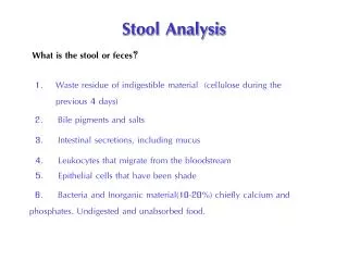
Stool analysis:
Stool analysis:. A stool analysis is a series of tests done on a stool (feces) sample for differential diagnosis of certain diseases of digestive system. Stool analysis procedure is divided into: 1-Physical Examination. 2-Chemical Examination. 3-Microscopic Examination.

Stool analysis:
E N D
Presentation Transcript
Stool analysis: A stool analysis is a series of tests done on a stool (feces) sample for differential diagnosis of certain diseases of digestive system. Stool analysis procedure is divided into: 1-Physical Examination. 2-Chemical Examination. 3-Microscopic Examination.
Clinical significance of stool analysis: 1-Diagnosis of digestive system infectious diseases: Bacteria, parasites, virus, and fungi. 2-Diagnosis of pancreas disorders (inflammation); which associated with malabsorption of nutrients. 3-Primary screening test for some types of digestive system malignancy such as: Colon cancer. 4-Primary screening test for peptic ulcer disease, and some types of anemia.
For whom Stool analysis is urgently required? 1-Patients with abdominal pain. 2-Patients with diarrhea. 3-Patients with anemia. Other situations by which Stool analysis is non-urgently required: 1-Patients who is too thin or do not grow well. 2-Patient with stool color that is changed to abnormal color.
Stool analysis: 1-Physical Examination: A-Color:Normal feces hasa dark brown color. (Bilirubin in the presence of bacteria will be oxidized to urobilinogenwhich give stool its color). Abnormal color: 1- Black color indicates blood of upper GIT origin(melena). 2- Red color indicates blood of lower GIT origin. 3- White color indicates yeast fermentation (Candida). -Very pale color indicates biliary obstruction or barium enema.
N Physical Examination: B-Consistency: -Normal feces is solid to semi-solid depending on diet. -A ribbon like fecal specimen could indicate irritable bowl syndrome or GIT obstruction. -Liquid stool indicates diarrhea (Gastroenteritis). -80-170 gm/day.
Chemical Examination: 1-The pH: The pH of the stool is 7.0-7.5. 2-Suger contents: The stool contains less than 0.25 grams per deciliter (g/dL) or less than 13.9 millimoles per liter (mmol/L) of sugars. -Elevated Suger indicates (more than 0.5g/dl): -Lactose intolerance. 3-Fat contents: The stool contains 2-7 grams of fat per 24 hours (g/24h).
N High levels of fat(steatorrhea) in the stool may be caused by diseases such as pancreatitis, celiacdisease(allergy to Gluten protein in wheat),or cysticfibrosis. 4-Occult blood:(The stool guaiac test): -Occult blood can be detected chemically : 1- Paper surface with phenolic compound alpha-guaiaconic acid. 2- Stool applied to the paper. 3-Hydrogenperoxide oxidizes alpha-guaiaconic acid to dark- blue color within two seconds. 4-Heme is a catalyst of this reaction.
N Patients should be instructed to avoid red meat, horse raddish, Asprin, Vitamin C as they interfere with the test by their catalases and peroxidases . Clinical significance of the test: 1-Diagnosis of Colorectal cancer. 2-Diagnosis of ulcer hemorrhoids. 3-Invasive Gastroenteritis.
Microscopic Examination: -Fecal leukocytes, especially neutrophils are associated with dysentery. -They can be detected by dried smears of the stool stained with gram stain. -Wet-mount smear(0.9% Saline) or Iodine stained smear should be prepared for parasites identification. 1-Wet-mount smear : Show the motility of active Protozoa. 2-Iodine smear: Show the nucleus and karyosome of protozoa.
Amoebic dysentery: Entamoeba histolytica(Rhizopoda): -Offensive stool. -Faecal matter mixed with blood and mucus. -RBCs, pus, mucus, and Entamoeba histolyticacyst or/and trophozoite (central karyosome). -Trophozoite show motility in one direction.
Giardia intestinalis : Gastrointestinal Mastigophora. Habitat:Small intestine especially in duodenum. Disease: Fatty diarrhea especially in children. Morphology: Trophozoite: four pairs of flagella. Giardia cyst (infective and diagnostic) Giardia trophozoite (diagnostic stage).
Balantidial dysentery: Balantidiumcoli: -Ciliophora. -Kidney-shaped Macronucleus. -Small micronucleus. -Ingestion of Contaminated pork meat.
Helminthes: Schistosoma mansoni: Trematoda. Intestinal bilharziasis. Diagnosis: finding of ova in stool. Ova with Lateral spine (diagnostic stage)
Cysticercosis: Taenia(Cestoda) infection: 1-Taeniasaginata(beef tapeworm). 2- Taeniasolium(pork tapeworm). Diagnostic stages: 1-Gravid segments. 2-Hexacantho- embryonated ova.
N Ascaris lumbricoides: -Nematoda. -Diagnostic stage: 1-Fertilized, un-fertilized ova. 2-Embryonated ova. 3-Adult stage.
N Hook worms: Ancylostomaduodenale. Nematoda. Diagnostic stage: 4-8 cell stage Embryonated ova.
N Enterobius vermicularis: -Nematoda. -Diagnostic stage: D-shaped ova. D-Shaped Ova.
"Scotch tape test" It is best done at night during the characteristic intense itching or early in the morning before any bathing or washing. Wrap a piece of cellophane tape around a tongue depressor, sticky side out, and press it to the skin around the anus to collect any eggs. Take the tape to a doctor, who will put it under a microscope to look for pinworm eggs. • The doctor may ask the person to use several pieces of tape to increase the likelihood of seeing the eggs. A single specimen will detect approximately 50% of cases; 90% of cases will be detected if the test is repeated three times. • Blood tests are not necessary to establish the diagnosis of pinworm infection. • The doctor may decide based upon the patient's symptoms that pinworms are present and may treat with medication without doing any tests.
N Strongyloidesstercoralis: -Free-living Nematoda. -Diagnostic stage: Rhabditiform larvae in stool.