Hematopoietic Cell Transplantation
540 likes | 585 Vues
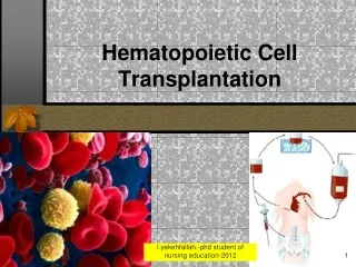
Hematopoietic Cell Transplantation. Definition. Hematopoietic cell transplantation (HCT) is a potentially curative treatment for malignant and nonmalignant diseases, including leukemia, lymphoma, multiple myeloma, aplastic anemia, hemoglobinopathies, and congenital immune deficiencies.

Hematopoietic Cell Transplantation
E N D
Presentation Transcript
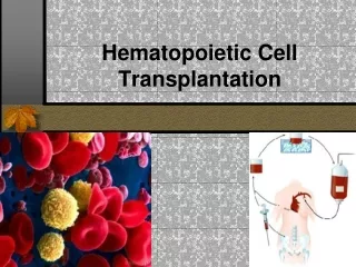
Definition • Hematopoietic cell transplantation (HCT) • is a potentially curative treatment for malignant and nonmalignant diseases, including leukemia, lymphoma, multiple myeloma, aplastic anemia, hemoglobinopathies, and congenital immune deficiencies. Dr.yekehfallah.-phd of nursing -2015
History of Bone Marrow Transplantation • First successful human bone marrow transplantation procedure (1968). Dr.yekehfallah.-phd of nursing -2015
Stem cells • Stem cells are ‘generic’cells that develop into particular types of cells. So they may become nerve cells, muscle cells, blood cells… in fact, any cell in the body! Stem cells divide over and over to produce new cells. Dr.yekehfallah.-phd of nursing -2015
Stem cells, bone marrow and blood cells One of the main places you find stem cells is in bone marrow. Stem cells in bone marrow produce new blood cells to replace those that have died. When the cells are mature they are released into the bloodstream. A ‘bone marrow’ donation is really a donation of stem cells. Bone marrow is found in the cavities inside the long and flat bones of the body. Dr.yekehfallah.-phd of nursing -2015
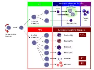
6 • Types of stem cells: • Pluripotent • Multipotent • Committed progenitor cells • Multipotent blood cells: • Common lymphoid • Common myeloid • Committed stem cell makes specific blood cells (CFU)–stimulated by erythropoietin, thrombopoietin, granulocyte-mononcyte CSF Dr.yekehfallah.-phd of nursing -2015
Leukocytes White blood cells Defend body through: the inflammatory process phagocytosis removal of cell debris immune reactions 7 Dr.yekehfallah.-phd of nursing -2015
White Blood Cell Types:Granulocytes and Agranulocytes Granulocytes–visible granules in the cytoplasm. Granules contain: Enzymes Other biochemicals that serve as signals and mediators of the inflammatory response 8 Dr.yekehfallah.-phd of nursing -2015
Granulocyte cell types: Neutrophils– phagocytes Eosinophils– red granules, associated with allergic response and parasitic worms Basophils– deep blue granules - Release heparin, histamine and serotonin 9 Dr.yekehfallah.-phd of nursing -2015
Agranulocytes Granules too small to be visible Monocytes– become macrophages Lymphocytes– B cells and T cells = immune functions 10 Dr.yekehfallah.-phd of nursing -2015
WBC’s originate in red bone marrow from stem cells. Granulocytes mature in the marrow and have a lifespan of hours to days Agranulocytes finishmaturing in blood, or in other locations. Monocytes live about 2 - 3 months, lymphocytes for years. Dr.yekehfallah.-phd of nursing -2015
Production of WBC’s increases in response to : Infection Presence of steroids Decreased reserve of leukocyte pool in bone marrow 12 Dr.yekehfallah.-phd of nursing -2015
13 Dr.yekehfallah.-phd of nursing -2015
14 Dr.yekehfallah.-phd of nursing -2015
Diseases Treated with Bone Marrow Transplantation • Acute leukemia (ALL, AML) • Chronic myelogenous leukemia • Aplastic Anemia • Myeloproliferative Disorders • Multiple Myeloma • Non-Hodgkin’s lymphoma • Hodgkin’s Disease • Chronic lymphocytic leukemia • Genetic Disorders (Thalassemia, others) • Solid tumors (RCC, neuroblastoma, germ cell tumors) • Congenital immunodeficiency diseases • Lymphomas • Metabolic disease of childhood • Myelodisplasia Dr.yekehfallah.-phd of nursing -2015
Stem cell (“bone marrow”) donation There are three ways to collect stem cells from a donor: • Bone marrowA donor has a small operation under general anesthetic. Marrow is harvested from the iliac crest under general anesthesia • Circulating bloodA donor’s circulating stem cells are boosted with a special drug. Then they are connected to a cell separator machine, which collects the stem cells and returns the rest of the blood to the donor. Growth factors are frequently used alone (e.g., granulocyte colony-stimulating factor, or G-CSF) or in combination with chemotherapy (in autologous HCT) for mobilization of hematopoietic stem cells, which are collected by blood leukapheresis. Mobilized†blood cells engraft faster than marrow-derived cells. • Cord bloodSelected hospitals offer new mothers the chance to donate the blood that remains in the placenta and umbilical cord after their baby’s birth. Collected from the umbilical cord after delivery of a baby. Engraftment takes longer compared with other sources of stem cells. Dr.yekehfallah.-phd of nursing -2015
Advantages / disadvantages for patients • Advantages: • Cord blood: • hasn’t been exposed to environment so less likely to contain viral infection; • requires less stringent matching; • once collected is banked and can be readily available at short notice. • Bone marrow and circulating blood: • tend to have a greater number of stem cells in the donation, so tend to be accepted into the patient’s body more quickly. • Disadvantages • Cord blood: • tends to have less stem cells in the donation. • Bone marrow and circulating blood: • finding a match and arranging a donation can take weeks, or months (this is time the patient may not have). Dr.yekehfallah.-phd of nursing -2015
Bone Marrow “Mobilized” peripheral blood stem cells Umbilical cord blood Cryopreserved Fresh SOURCE OF STEM CELLS Dr.yekehfallah.-phd of nursing -2015
Potential Donors • Self • HLA-matched sibling • HLA-mismatched sibling • Matched unrelated • Cord blood Dr.yekehfallah.-phd of nursing -2015
DonorLimitations • 25 – 30% of patients have an HLA-identical sibling. • Marrow procured from unrelated living donor • Marrow procured from related HLA-identical or HLA non –identical living donor • Autologous transolantation(marroe procured during remession) Dr.yekehfallah.-phd of nursing -2015
Stages of Allogeneic Transplantation Preparation Transplantation Recovery Immune suppression Disease treatment IV Infusion GVHD Prevention Infection control Nutrition Hematopoietic recovery Dr.yekehfallah.-phd of nursing -2015
RATIONALE FOR BMT • High dose chemotherapy (dose - response curve) • Allogeneic effect (graft-versus- tumour effect) • Replacement of abnormal stem cells (aplastic anaemia, thalassaemia, sickle cell disease, gene therapy etc) • Immunological effect (autoimmune disease, solid organ transplants) Dr.yekehfallah.-phd of nursing -2015
RISKS OF BONE MARROW TRANSPLANT • Short term (TRM) • Sepsis,AGVHD, multi-organ failure or toxic death • Longer term • Chronic graft-versus-host disease (lung,liver, skin) • Relapse • Infection • Endocrine Dr.yekehfallah.-phd of nursing -2015
Factors influencing survival • Disease factors • Remission, relapse, refractory • Transplant related mortality • Donor factors • Age, sex, conditioning • Recipient factors • Age, CMV status, performance status • Risk of graft-versus-host disease • Donor factors • PBSC, matching, age, parity • Recipient factors • Age Dr.yekehfallah.-phd of nursing -2015
GENERAL PRINCIPLES • Epidemiology. Current estimates of annual numbers of HCT are 45,000 to 50,000 worldwide. Approximately two thirds of patients have autologous HCT and one third have allogeneic HCT Dr.yekehfallah.-phd of nursing -2015
Risk factors • The likelihood of developing transplant-related complications depends on: 1/patient age 2/intensity of the preparative regimen 3/type and stage of the underlying disease 4/presence of comorbidities. 5/allogeneic HCT recipients have a greater risk of transplant-related morbidity and mortality than autologous HCT recipients 6/HLA disparity between donor and recipient further increases the risk owing to the greater likelihood of developing GVHD and graft rejection Dr.yekehfallah.-phd of nursing -2015
Prognosis Prognosis after HCT is highly variable and is influenced by numerous factors that predict for mortality related to the transplant procedure itself and to recurrent malignancy after surviving the transplant: - Patients with chronic myelogenous leukemia (CML) in chronic phase who have HCT from an HLA-identical sibling, for example, have a greater than 80% to 90% chance of long-term survival -In contrast, less than 50% of patients with more advanced leukemia at the time of HCT will be cured Dr.yekehfallah.-phd of nursing -2015
TransplantationProcedure Dr.yekehfallah.-phd of nursing -2015
Anesthesic Management • Intravenouse anesthesia sould be procured. • Intravenouse, Thiopental, Fentanyl ,Vecuronium can be used in common doses • Maintanance can be provide with Propofol and Isoflurane. Dr.yekehfallah.-phd of nursing -2015
Step 1: Bone marrow transplant with less toxic recipient treatment that includes antibodies.Donor marrow is T cell depleted Blood cells are a mixture of donor and host: Mixed chimerism is achieved without GVHR Wait 1-2 months. Inflammation from preparative treatment subsides. Step 2: Infuse donor T cells. Donor T cells interact with “presenting cells” of mixed chimera to maximize GVHR Donor T cells are armed to kill tumor cells that express recipient antigens. They stay inside the blood and lymph, where tumor is. T cells don’t go to skin/gut/liver. There is no GVHD. Tumor is killed Dr.yekehfallah.-phd of nursing -2015
TRANSPLANT RELATED COMPLICATIONS Dr.yekehfallah.-phd of nursing -2015
The problems after HCT that typically cause procedure-related morbidity and mortality can broadly be categorized into five groups: *hemolysis, • ** toxicity of the preparative regimen, ***infection, • **** bleeding, • ***** GVHD Dr.yekehfallah.-phd of nursing -2015
Hemolysis • General principles. • The inheritance of blood group antigens (e.g., ABO, Rh) is independent of that of the HLA antigens. Dr.yekehfallah.-phd of nursing -2015
Hemolysis • Etiology/pathophysiology: 1/ Acute (at the time of infusion) or delayed (5 to 15 days after HCT) immunohemolytic complications due to ABO incompatibility occur in patients with minor ABO-incompatibility. 2/ Immediate hemolysis occurs if the graft contains preexisting isohemagglutinins that lyse recipient RBCs. Dr.yekehfallah.-phd of nursing -2015
Hemolysis • Etiology/pathophysiology: 3/ Delayed hemolysis is due to generation of new isohemagglutinins by passenger lymphocytes in the graft. 4/Delayed hemolysis due to minor ABO incompatibility is a rare complication but can be dramatic and life-threatening. 5/ Major ABO incompatibility (recipient-derived isohemagglutinins directed against donor RBCs) may lead to chronic hemolysis and pure red cell anemia (PRCA). Dr.yekehfallah.-phd of nursing -2015
Hemolysis • Diagnosis 1/Clinical presentation: a/ Mild hemolysis due to major ABO incompatibility may be associated with prolonged RBC transfusion requirements . b/Delayed hemolysis due to minor ABO incompatibility may cause rapid lysis of all recipient RBCs over a few days. This may lead to acute renal failure or pulmonary edema and may be fatal. Plasma exchange in the recipient and plasma removal or RBC removal from the graft are standard procedures that minimize the risk of preventable hemolytic complication after ABO-mismatched HCT. Dr.yekehfallah.-phd of nursing -2015
Hemolysis • Diagnosis 2/Laboratory and radiologic studies: a/Emergence of donor-derived RBC and isohemagglutinin titers should be monitored after allogeneic HCT b/Serum levels of (indirect) bilirubin and lactate dehydrogenase (LDH), reticulocyte counts, and the direct agglutinin test (DAT) are useful markers of hemolysis Dr.yekehfallah.-phd of nursing -2015
Hemolysis Diagnosis 3/Differential diagnosis: a/Parvovirus 19 infection may cause PRCA after HCT. b/NonABO-related Coombs-positive autoimmune hemolytic anemia (AIHA) may develop late after HCT c/Drugs such as fluarabine or infections with mycoplasm may also produce hemolysis. Thrombotic microangiopathy (TM) is frequently related to cyclosporine or tacrolimus and associated with RBC fragmentation (schistocytes) d/Hyperbilirubinemia after HCT may have many other causes (e.g., sinusoidal obstruction syndrome [SOS] of the liver; drug effects) e/Dimethylsulfoxide (DMSO), a cryoprotectant used to store autologous stem cells, may cause anaphylactic and nonallergic reactions (e.g., hypotension, dyspnea, flushing, diarrhea) during infusion of thawed stem cell products. Dr.yekehfallah.-phd of nursing -2015
Hemolysis Treatment: 1/ Chronic hemolysis due to major ABO incompatibility is usually self-limited. 2/ Patients with refractory or more acute immune hemolysis may require interventions aimed at suppressing ongoing donor-directed isohemagglutinin production (corticosteroids, donor lymphocyte infusion, rituximab) or removal of the offending antibody (plasma exchange). 3/Supportive care measures to maintain renal function are critical Dr.yekehfallah.-phd of nursing -2015
Toxicity of the preparative regimen • Cytotoxic chemotherapy with or without total body irradiation (TBI) may compromise the function of the lungs, heart, kidneys, nervous system, and gastrointestinal tract including the liver. This type of toxicity occurs predominantly within the first 3 to 4 weeks after HCT. Dr.yekehfallah.-phd of nursing -2015
Infection General principles • Infections are frequent complications after autologous and allogeneic HCT. Their pattern of occurrence is determined by a number of factors, including the recipient's pretransplant history • and underlying disease, • the intensity of the preparative regimen, • the regimen used for infection prevention, the microbiological flora of the patient and of the individual transplant unit, • and the degree of immunosuppression after transplant . Dr.yekehfallah.-phd of nursing -2015
Bleeding Etiology/pathophysiology: Bleeding can occur as a result of thrombocytopenia or coagulopathy. Breakdown of mucosal barriers as a result of regimen-related toxicity and/or GVHD increases the likelihood of hemorrhage. CNS hemorrhage can be rapidly fatal. High-dose cyclophosphamide and BK virus infection are causes of hemorrhagic cystitis Dr.yekehfallah.-phd of nursing -2015
Bleeding Diagnosis: Oropharyngeal bleeding and epistaxis are obvious. Endoscopic evaluation (colonoscopy, esophagogastroduodenoscopy, bronchoscopy) or CNS imaging may be indicated according to the clinical picture Dr.yekehfallah.-phd of nursing -2015
Bleeding Treatment: A platelet count below 10,000/mm3 increases the risk of spontaneous bleeding and should be treated prophylactically with platelet transfusion. The decision to transfuse platelets above the prophylactic threshold of 10,000/mm3 should be guided by the clinical situation. Patients who have received multiple transfusions can become alloimmunized and demonstrate poor response to platelet transfusion. Transfusion of unpooled (single-donor) or HLA-matched platelets may be helpful. Bleeding can be seen on bronchoscopy. The treatment consists of high doses of corticosteroids (e.g., 1 g per day for 3 days). Dr.yekehfallah.-phd of nursing -2015
Graft-versus-host disease General principles GVHD is a major cause of morbidity and mortality after allogeneic HCT. Patients who develop GVHD, however, have lower relapse rates than patients without GVHD, which can be explained by an immunologic graft-versus-tumor effect that helps eradicate the underlying malignancy Dr.yekehfallah.-phd of nursing -2015
Graft-versus-host disease Etiology/pathophysiology The GVHD syndrome is caused by donor T cells that are activated by immunologically disparate HLA or non-HLA antigens in the recipient, resulting in an inflammatory immune response. Older patient age, HCT from a mismatched and/or unrelated donor, and a female donor increase the risk of developing GVHD Dr.yekehfallah.-phd of nursing -2015
Graft-versus-host disease Diagnosis Clinical presentation: Acute GVHD (defined as occurring before posttransplant day 100) tends to have a more sudden onset and may involve the skin, gastrointestinal tract, and liver. Signs and symptoms include an erythematous rash, nausea, vomiting, diarrhea, and liver function abnormalities. Clinical features of chronic GVHD (defined as occurring after posttransplant day 100), which typically has a more insidious onset, may involve the skin, eyes, joints, and liver and are reminiscent of autoimmune diseases such as systemic sclerosis. Dr.yekehfallah.-phd of nursing -2015
Graft-versus-host disease • Laboratory and radiologic studies: Skin punch biopsy, gastrointestinal endoscopy with biopsy, and liver biopsy confirm the diagnosis in the appropriate clinical setting. Cholestatic jaundice is the hallmark of liver involvement. Pulmonary function test and lung biopsy in patients with presumed BOOP to rule out infectious etiologies. Dr.yekehfallah.-phd of nursing -2015
Graft-versus-host disease Treatment: Despite prophylaxis against GVHD (e.g., cyclosporine plus methotrexate), 40% to 80% of allogeneic HCT recipients develop this complication. Corticosteroids are standard first-line therapy of GVHD (e.g., prednisone 2 mg/kg per day; slow taper after 2 weeks). Dr.yekehfallah.-phd of nursing -2015