Download
1 / 87
870 likes | 1.18k Vues
Unit 1 The Power of Reproduction. Chapter 1 – The Cell Cycle and Asexual Reproduction Amazing Cells. Animal Cell. Plant Cell. 1.1 The Cell (Pages 4 – 16).

E N D
Chapter 1 – The Cell Cycle and Asexual ReproductionAmazing Cells
Animal Cell Plant Cell
1.1 The Cell (Pages 4 – 16) Scientists have long been fascinated with life. After thousands of years of studying it we still haven’t figured out how it works. Some history: Assignment: The Cell Question Sheet Label Cell Diagrams (1-6 & 1-7) Cell Structures Chart
Chapter 1.1 – The Cell Question Sheet Record the date and contribution to cell theory for the following scientists: (22) Aristotle (384-322 BC) – animal and plant kingdom (animals higher, spontaneous generation) Zacharias Janssen (1590) – first compound microscope Robert Hooke (1665) – microscope – referred to cells as “rooms”
Chapter 1.1 – The Cell Question Sheet Record the date and contribution to cell theory for the following scientists: (22) John Ray (1667) – “species” organism that reproduce with their own kind Francesco Redi (1668) – disproved spontaneous generation (flies and maggots)
The following photomicrographs were taken through a Leeuwenhoek microscope.
Anton van Leeuwenhoek (1632 – 1723) - microscope Robert Brown - nucleus is part of the cell Schleiden/Schwann (1838/1839) – all organisms are made of cells Alexander Carl Heinrich Braun (1845) – the cell is the basic unit of life
Charles Darwin and Alfred Wallace – species form variations Rudolph Virchow (1821 – 1902) – cells come from cells Louis Pasteur (1860) – disproved spontaneous generation at the microscopic level (life comes from life)
Like all sciences, biology has its own set of tools, techniques, and investigative methods. What tools helped scientists develop the cell theory? (3) What methods helped scientists develop the cell theory? (2)
What methods helped scientists develop the cell theory? (2) State a question Form a hypothesis (educated guess – what you think will happen) Design an experiment Make observations Draw conclusions – answer hypothesis
The internet was originally designed to allow scientists all over the world to communicate quickly and easily with each other. What invention had a similar effect on scientific communication in earlier centuries? (1)
What can you conclude about the relationships between scientific discovery, tool inventions, and new methods? (1)
Cell theory states that: (4) all living organisms are made up of one or more cells Cells are the basic unit of life Cells come from cells Activity of entire organism depends on total activity of its independent cells
Assignment BLM 1-5, 1-8, 1-9, 1-10, Investigation 1-B
Cell Structures Chart Cell Size and Scale Nucleus contains the cell’s genetic material holds instructions for making/building the cell Nuclear Membrane encloses the cells’ genetic material contains the DNA
Cell Structures Chart DNA Deoxyribonucleic Acid Chromatin Long strands of DNA Nucleolus dark area within the nucleus manufactures ribosome parts
Cell Structures Chart Ribosomes contribute to the manufacture of proteins Cell Membrane separates the contents of the cell from its surroundings selectively permeable membrane that allows only certain material in and out of the cell
Cell Structures Chart Cytoplasm jelly-like material supports the nucleus and other organelles Endoplasmic Reticulum a folded membrane that forms a series of canals transports materials to different parts of the cell (like our circulatory system)
Cell Structures Chart Mitochondria transform energy for the cell (sugar to ATP) cellular respiration takes place here (this is not in your text, please copy it) Golgi Bodies package useful materials then it secretes them to the outside of the cell (like our digestive system)
Cell Structures Chart Vacuoles fluid filled store houses for water, waste and nutrients (like our digestive system) Lysosomes break down food and digest wastes and worn out cell parts (like our digestive system)
Cell Structures ChartPlants Only Cell Wall Provides structure and support for the cell (acts like the cells’ skeleton) Chloroplasts this is where photosynthesis takes place photo means “involving light” and “synthesis means “to make”
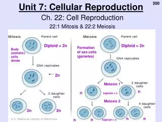
1.2 Understanding the Cell Cycle Cells grow and then divide to make new cells. New cells are used to replace dead ones OR for growth of the organism. YOU started out as 1 cell! Before a cell can divide into 2 cells it must produce almost twice as many organelles (replication). A cell’s stages of life are called phases.
1. Interphase Most of cell’s life DNA in thin strands called Chromatin replicate. Chromatin coils up to form double stranded Chromosomes. A Centromere connects the original chromatin with its identical replicate. The cell has a complete extra copy of DNA.
2. Prophase Duplicate DNA is easily seen under microscope. Nucleolus and Nuclear Membrane disappear. Centrioles move to opposite sides of the cell. (Fishing boats) Spindle fibres (like a scaffold) grow out of each centriole and attach to centromere. (Fishing line)
3. Metaphase Spindle fibres pull on centromeres Chromosomes move to line up in the middle. 4. Anaphase Spindle fibres shorten and pull centromere apart. One copy of DNA goes to each side.
5. Telophase A complete set of chromosomes arrives at each centriole. Spindle fibres disappear. Nuclear membrane and nucleolus reform. Chromosomes uncoil into thin chromatin.
5. Telophase (cont’d) Cell membrane pinches together in the middle (cytokinesis) Two cells form (animal cells). A cell plate grows across the middle of the cell forming a new cell wall between the two cells. (plants)
Mitosis Animation Clips http://www.pbs.org/wgbh/nova/miracle/divide.html# http://www.johnkyrk.com/mitosis.html organelles and mitosis In depth mitosis http://www.dnatube.com/video/2380/Interpretive-Mitosis
Assignment BLM’s 1-14, 1-15, 1-16 & 1-17 Mitosis Crossword & Mitosis Chart Mitosis Quiz
1.3 The Cell Cycle in YOUR Body Some common chromosome counts:
Cell need to divide for the following reasons: Normal Cell Replacement Cells die of old age and need to be replaced. See pg 25 for cell life spans. About 3 billion cells die in your body every minute. Cells die due to damage or when they don’t get enough food or oxygen. Regeneration - Healing of damaged tissue or the replacement of body parts is called regeneration. (NOTE: see note under Figure 1.17 – pg 26)
Growth As organisms grow larger, their cells stay the same size They just get more of them.
Aging is connected to the slowing of the cell cycle. The cells do not divide as often or as quickly. Aging
Cancer 1. Caused when the DNA of a cell becomes damaged by: • Tobacco • Asbestos • Certain chemicals • some viruses • Radioactivity • UV radiation
The damage injures but does not kill the cell. The cell no longer functions properly and the DNA no longer has correct information about when and how quickly to divide. The result: Useless cells divide often and quickly forming a lump. If the cancer cells can easily be transported the cancer can spread all over. The lump crowds out good cells and use up a lot of food and oxygen. Worksheet & Cancer Research
Explain a process in the human body in which there is evidence of the cell cycle at work. (1) Growth and development