Download
1 / 25
300 likes | 879 Vues
INFECTIONS OF THE UPPER RESPIRATORY TRACT. Elizabeth Wasserman Clinical Microbiologist, Pathcare Laboratories Extraordinary professor, Division of Medical Microbiology, Stellenbosch University. RESPIRATORY INFECTIONS. Divided into: Upper respiratory tract infections

E N D
INFECTIONS OF THE UPPER RESPIRATORY TRACT Elizabeth Wasserman Clinical Microbiologist, Pathcare Laboratories Extraordinary professor, Division of Medical Microbiology, Stellenbosch University
RESPIRATORY INFECTIONS Divided into: • Upper respiratory tract infections • Lower respiratory tract infections Division is the larynx. Normal flora is found above the larynx, while the environment below the larynx is sterile under normal conditions.
Normal flora of the upper respiratory tract 1. Commonly carried bacteria: Streptococcus viridansNeisseriaspp. DiphtheroidsAnaerobiesecocci, fusiforme & Bacteroides2. Bacterial pathogens that can be carried asymptomatically: Streptococcus pyogenes Streptococcus pneumoniaeHaemophilusinfluenzaeCorynebacteriumdiphtheriae Moraxellacatarrhalis3. Organisms associated with colonization as a result of antimicrobial therapy: Coliforms - Klebsiella spp., E. coli, etc. Pseudomonas spp. Candida albicans
Incidence of upper airway infections: • Viral infections in pre-school children – up to 6 x per year. • Bacterial infections are also very common in children.
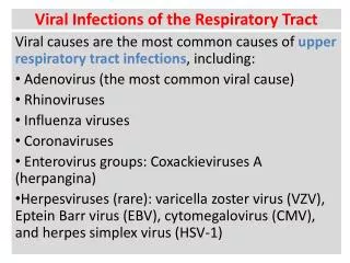
"Common cold"(Coryza) Ethiology: - Corona virus - R S V - Para-influenza virus - Coxsackie A21 and B3 - Echovirus 11 + 20 - Adenoviruses
Ethiology: Viruses: - Adenovirus - EBV - Enterovirus - Prodrome of for example measles - CMV - Herpes simplex
Bacteria: - S. pyogenes including Lancefield groups C + G - C. diphtheriae and C. ulcerans (rare) - Arcanobacterium haemolyticum – especially in adults - Vincent’s organisms (B. vincenti + anaerobe fusiform basilli) - T. pallidum (2o syphilis). - N. gonorrhoeae
Laboratory diagnosis not necessary as routine • Treatment usually empiric with narrow spectrum beta lactam drug
Complications of streptococcal infection: Direct: - peritonsillarabscess - Otitis media - scarlet fever Indirect: - Rheumatic fever -Acute Glomerulonephritis
Otitis media Ethiology: Viruses (50%) Bacteria: - S. pneumoniae - H. influenzae - S. pyogenes - S. aureus - M. catarrhalis - M. pneumoniae (rare)
Laboratory diagnosis: Specimen collection underdirectvission, preferably an aspirate: Gram stain culture http://www.rnceus.com/otitis/images/tympanocentesis.jpg
Treatment: - Amoxicillin - beta-lactamase producing organisms (H. influenzae + M. catarrhalis): Augmentin (Amoxicillin + Clavulanic acid) or 2nd generation cephalosporin Complicactions: - Chronic suppurative otitis media - Mastoiditis - secretory otitis media ("gum ear")
Otitisexterna Clinical presentation: Irritation and secretion of the external ear. Etiologic: Bacterial: - S. aureus - Proteus spp - P. aeruginosa("Malign Otitisexsterna“) Fungi: - Aspergillusniger - C. albicans
Laboratory diagnosis: Culture of pus /swab. Treatment: - Ear toilet - Topical antibiotics according to sensitivities
Sinusitis Etiologic: Viruses Bacterial: - H. influenzae - S. pneumoniae - Anaerobe and micro-aerophilic streptococci - S. aureus • S. pyogenes • M. catarrhalis
Treatment: - Empiric therapy amoxicillin. Beware of beta-lactamase producing organisms: Augmentin, 2nd generation cephalosporin. - Allergic patients: cotrimoxazole, anaerobic cover - metronidazole. Complications: - Osteomyelitis - Meningitis - cerebral abscess
Ethiology: Viral: - parainfluenza 1,2,3 - Influenza A - Rhinoviruses, RSV Bacterial: - M. pneumoniae
Epiglottitis Ethiology: - H. influenzae type b Diagnosis: - Examine only in theatre - Cherry red epiglottis - XR picture - Blood cultures
Pertussis • Caused by the bacterium Bordetella pertussis • Limited to the human host and transmitted from person to person by droplet spread • Severe disease of childhood • Organisms adhere to the ciliated epithelial cells of the respiratory tract and start to multiply. Not invasive. • Cause inflammation and damage to the ciliated epithelial cells by means of various toxins (pertussis toxin, endotoxin, trageal cytotoxin, adenylate cyclase toxin)
Clinical characteristics • Incubation period 1 – 3 weeks • Starts with a catarrhal phase • One week later: dry cough that becomes paroxysmal (bouts of coughing) • Paroxysmal cough followed by a ‘whoop’. • Superfluous phlegm production • In spite of the severity of the disease, the symptoms are limited to the respiratory tract.
Complications • CNS: anoxia, exhaustion • Secondary pneumonia Diagnosis • Clinical • Organisms isolated on special media that has to be inoculated at the bedside, from a throat swab or so-called ‘cough plates’ • PCR
Treatment • Supportive • Erythromycin: this may limit the severity and duration of the disease • It may also limit infectivity and the risk of secondary infections Prevention • Active immunization • Whole cell vaccine • In the future: ?subunit vaccine • Erythromycin prophylaxis for high risk, close contacts.