Materials used in the different studies
Reevaluation ?. 1) Analyze and assay 4 different HDL preparations using Gel Filtration. 2). Remove HDL particles from HDL prep. using anti. -. ApoAI. immunoadsorbtion. . 3). Correlate APC / . ProS. anticoagulant response with HDL levels in plasmas.

Materials used in the different studies
E N D
Presentation Transcript
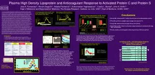
Reevaluation ? 1) Analyze and assay 4 different HDL preparations using Gel Filtration. 2) Remove HDL particles from HDL prep. using anti - ApoAI immunoadsorbtion . 3) Correlate APC / ProS anticoagulant response with HDL levels in plasmas. Plasma High Density Lipoprotein and Anticoagulant Response to Activated Protein C and Protein S Jose A. Fernandez1), Hiroshi Deguchi1), Natalie Pecheniuk1), Subramanian Yegneswaran1), Carole L. Banka2), John H. Griffin1) Dept of Molecular and Experimental Medicine, The Scripps Research Institute, La Jolla, USA1), Dept of Medicine, UCSD, USA2) #2249 Previously we reported that plasma high density lipoprotein (HDL) enhances activated protein C (APC)/protein S (PS) anticoagulant action in plasma clotting and factor Va inactivation assays (APC/PS enhancement) and that lower HDL levels are found in male venous thrombosis patients or in patients with recurrent venous thrombosis versus controls, giving rise to our hypothesis that HDL helps protect against venous thrombosis. In this study, we sought (1) to identify which HDL particles enhance APC/PS activity, (2) to assess correlations between this activity and HDL particle size, and (3) to evaluate the recent challenge to our HDL anticoagulant activity hypothesis (Oslakovic et al, J Clin Invest, 2010). To identify HDL subfractions with APC/PS enhancing activity, we subfractionated HDL by sequential density gradient ultracentrifugation over the HDL density range of 1.063-1.21 mg/dl. When the anticoagulant property of these HDL subtractions was studied, we found that the less dense HDL subfractions corresponding to HDL2 particles enhanced APC/PS anticoagulant activity much more than other HDL fractions. Thus, we identified larger HDL particles as the key subfraction for this anticoagulant property of HDL. Hence, we hypothesized that plasma levels of large HDL particles would correlate with plasma sensitivity of APC/protein S. To test this hypothesis, we used proton NMR to quantify the levels of large, medium and small HDL particles and to determine the average size of HDL particles in plasmas from 39 normal adults. We performed dilute tissue factor-induced clotting assays in the absence or presence of APC/protein S to define APC/PS sensitivity and assessed correlations. There was a positive correlation between large HDL particle concentration and plasma sensitivity to APC/protein S (r=0.40, p=0.02). The size of HDL particles was also positively correlated with plasma sensitivity to APC/protein S (r=0.42, p=0.01). As previously reported, apoAI concentrations which is major apolipoprotein in HDL correlated with plasma sensitivity to APC/protein S (r=0.52, p=0.0007). Thus, as hypothesized, apoAI-containing large HDL particle concentrations in plasma correlate very well with the anticoagulant response to APC/PS. Recently Oslakovic et al purported to show that APC/PS enhancement was not an intrinsic property of HDL and claimed that this activity was due to negatively charged phospholipid contaminants of HDL based on gel filtration (Superose 6) analysis of their frozen HDL preparation. Therefore, we applied Superose 6 gel filtration analyses to four different fresh, never-frozen HDL preparations coming either from commercial sources or from in-house preparations. Our studies showed that when each HDL prep was subjected to Superose 6 fractionation, the column fractions containing large HDL (apoAI positive fractions) enhanced APC/protein S anticoagulant activity in plasma clotting assays and also for the inactivation of factor Va in purified systems. Moreover, immobilized anti-apoAI-antibodies removed the APC/PS enhancing effect, further establishing the fact that apoAI-containing HDL particles enhance APC/PS activity. When we analyzed HDL preps stored at 4 °C over successive weeks, we found that HDL fractions lost the ability to enhance APC/PS, showing the relatively labile nature of this activity. Freezing and thawing HDL was also deleterious for this activity. Thus, the APC/PS enhancing activity of fresh, never-frozen HDL preps is primarily due to HDL particles. All of our new findings confirm our previous conclusion that HDL enhances APC/PS anticoagulant actions. Our extensive data set strongly contradicts the conclusions from the recent HDL study by Oslakovic et al who unfortunately performed their bioassays using frozen HDL made from previously frozen lipidmic plasma. In summary, freshly purified, non-frozen large HDL particles made from fresh plasma enhance APC/PS activity. The sensitivity of plasma of healthy adults to APC/PS anticoagulant effects is significantly correlated with the plasma levels of large HDL particles, suggesting the physiological importance of large HDL particles as enhancing the anticoagulant APC/PS system and supporting our hypothesis that large HDL particles may be protective against venous thrombosis, at least in part, via this activity. Conclusions To define the active molecular species in HDL, immobilized antibodies against apoA-I, were tested for their ability to absorb the anticoagulant activity of superpose 6 fractions. Anti–apoA-I IgG adsorbed 86% of the anticoagulant activity, whereas no significant (<5%) adsorption was observed for the control Sepharose beads. Thus, HDL fractions containing apoA-I provided the anticoagulant activity observed. Materials used in the different studies The fractions were tested for their ability to enhance FVa inactivation and prothrombin activation. The inactivation of purified coagulation FVa by APC and its cofactor, protein S, was studied using a two-step procedure. First, FVa was incubated with HDL fractions or buffer, APC, and protein S to inactivate FVa. Second, residual FVa activity was quantitated using prothrombinase assays. The pool of fractions corresponding to apo AI peak markedly enhanced APC-dependent loss of FVa. The fractions on the void volume also show inhibition although less potent. Fractions in the void volume were also found to stimulate prothrombin activation, whereas the ApoAI containing fractions did not stimulated prothrombin activation. Thus, the anticoagulant effects that were observed in HDL preparations isolated by ultracentrifugation were associated with the apoAI fractions. Superose 6 Chromatography Profile of HDL Prep’s When the anticoagulant property of these HDL subtractions was studied, we found that the lightest HDL subfractions were more anticoagulantly active as APC-cofactors than other HDL fractions. These HDL subfractions with anticoagulant activity correspond to the peak of phospholipid, cholesterol and protein in HDL2 containing large HDL particles.