Chapter 40 Maintaining the Internal Environment
Chapter 40 Maintaining the Internal Environment. 40.1 Truth in a Test Tube. Kidneys rid the body of excess water, excess or harmful solutes, and drugs Physicians routinely check urine to monitor their patient ’ s health


Chapter 40 Maintaining the Internal Environment
E N D
Presentation Transcript
40.1 Truth in a Test Tube • Kidneys rid the body of excess water, excess or harmful solutes, and drugs • Physicians routinely check urine to monitor their patient’s health • Athletic associations use urine tests to check for performance-enhancing substances as well as “street drugs”
40.2 Regulating Fluid Volume and Composition • All animals constantly acquire and lose water and solutes, and produce metabolic wastes • Excretory organs keep the volume and the composition of their internal environment – the extracellular fluid – stable • Metabolic wastes, particularly carbon dioxide and ammonia, affect the composition of the extracellular fluid
Products of Protein Breakdown A amino group B ammonia C uric acid D urea
How Invertebrates Maintain Fluid Balance • Marine invertebrates usually have body fluids with the same solute concentration as seawater – osmosis produces no net movement of water into or out of the body • In planarian flatworms and other freshwater animals, body fluids have a higher solute concentration than the surrounding water, so water enters by osmosis
Flatworms and Earthworms • Flatworms and earthworms have tubular excretory organs that deliver fluid with dissolved ammonia to a pore at the body surface • Flatworms have protonephridia with ciliated flame cells • Earthworms have nephridia that collect coelomic fluid
Planarian Protonephridia nucleus of flame cell cilia of flame cell spaces through which water enters tube pore at body surface
Earthworm Nephridia storage bladder loops where exchanges with blood in adjacent vessels occur funnel that collects coelomic fluid pore through which waste exits
Arthropods • Insects convert ammonia to uric acidcrystals, which Malpighian tubulesdeliver to the gut • Excreting uric acid rather than ammonia reduces water loss
Malpighian Tubules Malpighian tubule part of gut
Fluid Regulation in Vertebrates • Vertebrates have a urinary systemthat filters water, metabolic wastes and toxins out of the blood, and reclaims water and certain solutes • All vertebrates have paired kidneys – excretory organs that filter blood and adjust the level of solutes
Fluid Balance in Fishes • Bony fishes have body fluid that is saltier than freshwater, but less salty than seawater • Marine fish drink water, pump excess salt out through gills, and produce small amounts of urine • Freshwater fish do not drink, and produce large amounts of dilute urine
Marine Bony Fishes water loss by osmosis gulps water water loss in very small volume of concentrated urine cells in gills pump solutes out A Marine bony fish with body fluids less salty than the surrounding water; the fish is hypotonic relative to its environment.
Freshwater Bony Fishes water gain by osmosis does not drink water cells in gills pump solutes in water loss in large volume of dilute urine B Freshwater bony fish with body fluids saltier than the surrounding water; the fish is hypertonic relative to its environment.
Land Vertebrates • Waterproof skin and highly efficient kidneys help adapt amniotes to land • Reptiles and birds conserve water by converting ammonia to uric acid • Mammals excrete urea, which requires 20 times more water to excrete than uric acid • Mammals with limited access to fresh water tend to have large kidneys for their size and lose little water in their urine
Take-Home Message: Volume and composition of body fluid • All animals must rid the body of waste carbon dioxide and ammonia; many convert ammonia to urea or uric acid before excreting it. • Most animals have excretory organs that interact with a circulatory system to remove wastes from the blood and excrete them.
Take-Home Message: (cont.) • Invertebrate excretory organs include the ammonia-excreting nephridia of earthworms and the uric acid–excreting Malpighian tubules of insects. • All vertebrates have two kidneys. The volume of urine and the type of nitrogen-containing wastes excreted (ammonia, urea, or uric acid) vary among groups
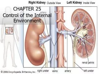
40.3 The Human Urinary System • Kidneys filter blood and form urine: • Fibrous outer layer (renal capsule) • Two inner zones: renal cortexand renal medulla • The body reclaims most of the filtrate; urine flows through ureters into a bladder that stores it • Urine flows out of the body through the urethra
heart Kidney (one of a pair) Blood-filtering organ; filters water, all solutes except proteins from blood; reclaims only amounts body requires, excretes rest as urine diaphragm adrenal gland Ureter (one of a pair) Channel for urine flow from one kidney to urinary bladder abdominal aorta inferior vena cava Urinary Bladder Stretchable urine storage container Urethra Urine flow channel between urinary bladder and body surface Figure 40-8a p718
renal cortex renal medulla (back of body) backbone right kidney left kidney renal artery peritoneum abdominal cavity renal vein (front of body) renal pelvis renal capsule ureter Figure 40-8bc p718
ANIMATED FIGURE: Human urinary system To play movie you must be in Slide Show Mode PC Users: Please wait for content to load, then click to play Mac Users: CLICK HERE
Nephron Structure • Nephron • A microscopic tube with a wall one cell thick • Begins in the cortex, where it folds to form a cup-shaped Bowman’s capsule • Enters the medulla as a proximal tubule, turns at the loop of Henle, reenters the cortex as a distal tubulewhich drains into a collecting duct
Blood Vessels Around Nephrons • Renal arteries branch into afferent arterioles, which branch into a capillary bed (glomerulus) inside a Bowman’s capsule, which filters blood • Efferent arterioles branch into peritubular capillariesaround the nephron, which converge into venules and veins leaving the kidney
A Nephrons extend from the cortex into the medulla. Figure 40-9a p719
Bowman’s capsule (red) proximal tubule (orange) distal tubule (brown) Renal Cortex Renal Medulla loop of Henle (yellow) collecting tubule (tan) B Tubular portion of one nephron, cutaway view. The tubule starts at Bowman’s capsule. Figure 40-9b p719
efferent arteriole glomerulus afferent arteriole renal artery renal vein peritubular capillaries C Blood vessels associated with the nephron. The glomerulus is a ball of capillaries that have unusually leaky walls. Figure 40-9c p719
ANIMATED FIGURE: Human kidney To play movie you must be in Slide Show Mode PC Users: Please wait for content to load, then click to play Mac Users: CLICK HERE
Take-Home Message: Components of the human urinary system • The human urinary system has two kidneys, two ureters, a urinary bladder, and a urethra. Kidneys filter the blood and form urine. Urine flows out of the kidney through ureters, and into a hollow, muscular bladder. When the bladder contracts, urine flows out of the body through the urethra. • The functional unit of the kidneys is the nephron, a microscopic tubule that interacts with two systems of capillaries to filter blood and form urine.
ANIMATION: Tubular reabsorption To play movie you must be in Slide Show Mode PC Users: Please wait for content to load, then click to play Mac Users: CLICK HERE
40.4 How Urine Forms • Urine consists of water and solutes filtered from blood and not returned to it, plus unwanted solutes secreted from blood into the nephron’s tubular regions • Urine forms by three physiological processes: glomerular filtration, tubular reabsorption, and tubular secretion
Glomerular Filtration • Glomerular filtration is the first step in urine formation • Occurs at glomerular capillaries in Bowman’s capsule • The force of the heartbeat drives protein-free plasma out of glomerular capillaries and into the nephron’s tubular portion as filtrate
glomerulus inside Bowman’s capsule Figure 40-10a p720
glomerulus inside Bowman’s capsule outer wall of Bowman’s capsule afferent arteriole (from renal artery) filtrate (to proximal tubule) efferent arteriole (to peritubular capillaries) Figure 40-10b p720
Tubular Reabsorption • Tubular reabsorption returns most water and solutes to the blood • Occurs all along a nephron’s tubular parts • Nearly all water and solutes that leave the blood as filtrate later leave the tubule and return to the blood in peritubular capillaries
Tubular Secretion • Tubular secretion is the movement of substances from the blood in peritubular capillaries into the filtrate • Starts at the proximal tubule and continues all along a nephron’s tubular parts • Urine forms from water and solutes that remain in the tubule, and solutes secreted into the tubule along its length
Concentrating the Urine • Concentration of urine flowing down through the loop of Henle sets up a solute concentration gradient in surrounding interstitial fluid of the renal medulla • This gradient allows urine to become concentrated as it flows through the collecting duct to the renal pelvis • The body can adjust how much water is reabsorbed at distal tubules and collecting tubules
Urine Formation collecting tubule Bowman’s capsule distal tubule proximal tubule protein-free plasma Na+, Cl–, K+, nutrients, H2O H+ Na+, Cl–, H2O H+, K+ 3 1 2 Glomerular filtration Tubular secretion Tubular reabsorption 4 Na+ peritubular capillary H2O renal cortex renal medulla descending arm of loop of Henle ascending arm of loop of Henle H2O Na+ urea increasing solute concentration in interstitial fluid H2O urine to renal pelvis
ANIMATED FIGURE: Urine formation To play movie you must be in Slide Show Mode PC Users: Please wait for content to load, then click to play Mac Users: CLICK HERE
Take-Home Message:How is urine formed and concentrated? • During glomerular filtration, pressure generated by the beating heart drives water and solutes out of glomerular capillaries and into kidney tubules. • In tubular reabsorption, water, some ions, glucose, and other solutes move out of the filtrate and return to the blood in peritubular capillaries. • In tubular secretion, active transport proteins move solutes such as H+ and K+ from peritubular capillaries into the nephron for excretion.
Take-Home Message:How is urine formed and concentrated? • Differential permeability of the two arms of the loop of Henle sets up a concentration gradient in the interstitial fluid that draws water out of the collecting tubule. • Urine concentration depends on how much water flows out of the distal tubule and the collecting tubule. Hormones affect the concentration of solutes in urine by their effects on the permeability of these tubules.
40.5 Regulating Thirst and Urine Concentration • When you don’t drink enough water, a region of the hypothalamus (thirst center) notifies your cerebral cortex that you need to search for water • Hormonal controls act to conserve water already in the body
Effects of Antidiuretic Hormone • Antidiuretic hormone(ADH) • Released by the pituitary when sodium levels rise • Increases water reabsorption by stimulating insertion of aquaporins into plasma membranes of distal tubules and collecting ducts • Concentrates urine
Feedback Control of ADH Secretion hypothalamus ADH alert! Stimulus Response 5 1 2 pituitary gland 3 4
Effects of Aldosterone • Aldosterone • Released by the adrenal cortex • Increases salt reabsorption in collection ducts; water follows by osmosis; urine is concentrated • Decrease in volume of extracellular fluid stimulates arterioles in nephrons to release renin • Renin converts angiotensinogen to angiotensin I, converted to angiotensin II, which acts on the adrenal cortex to secrete aldosterone
Angiotensinogen is converted to Angiotensin I is converted to Angiotensin II encourages secretion of Aldosterone acts on kidneys to increase Na+ reabsorption Figure 40-13a p723
Effects of Atrial Natriuretic Peptide • Atrial natriuretic peptide (ANP) • Released by muscle cells in the heart’s atria when high blood volume causes walls to stretch • Directly inhibits secretion of aldosterone by acting on adrenal cortex • Indirectly inhibits secretion of aldosterone by inhibiting renin release • Increases glomerular filtration rate, makes urine more dilute
Take-Home Message: How do hormones affect urine concentration? • Antidiuretic hormone released by the pituitary causes an increase in water reabsorption. It concentrates the urine. • Aldosterone released by the adrenal cortex increases salt reabsorption, and water follows. It concentrates the urine. • Atrial natriuretic peptide released by the heart makes urine more dilute by discouraging secretion of aldosterone and increasing the rate of glomerular filtration.
40.6 Acid–Base Balance • Normal pH of extracellular fluid is 7.35 to 7.45 • Kidneys, buffering systems, and respiratory system work together to maintain the acid-base balance(H+ concentration) within a tight range • Kidneys are the only organs that can selectively rid the body of H+ ions