Maintaining the Internal Environment
Maintaining the Internal Environment. Impacts, Issues Truth in a Test Tube. Kidneys rid the body of excess water, excess or harmful solutes, and drugs – physicians routinely check urine to monitor their patient’s health. Maintenance of Extracellular Fluid.

Maintaining the Internal Environment
E N D
Presentation Transcript
Impacts, IssuesTruth in a Test Tube • Kidneys rid the body of excess water, excess or harmful solutes, and drugs – physicians routinely check urine to monitor their patient’s health
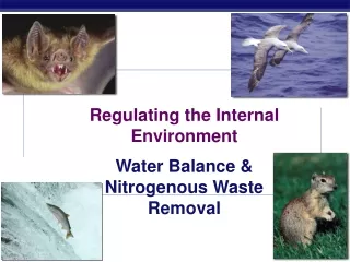
Maintenance of Extracellular Fluid • All animals constantly acquire and lose water and solutes, and produce metabolic wastes • Excretory organs keep the volume and the composition of their internal environment – the extracellular fluid – stable
plasma lymph, cerebrospinal fluid, mucus, and other fluids interstitial fluid Extracellular Fluid (ECF) (15 liters) Intracellular Fluid (28 liters) Human Body Fluids (43 liters) Fig. 41-2, p. 722
How Do Invertebrates Maintain Fluid Balance? • Sponges • Simple animals with no excretory organs • Wastes diffuse out across the body wall • Excess water is expelled by contractile vacuoles
How Invertebrates Maintain Fluid Balance • Flatworms and earthworms • Tubular excretory organs deliver fluid with dissolved ammonia to a pore at the body surface • Flame cells in flatworms • Nephridia in earthworms
Planarian Flame Cells nucleus cilia pair of highly branched tubules that adjust water and solute levels in body fluid filters through membrane folds flame cell opening at body surface Fig. 41-3, p. 722
body wall storage bladder loops where blood vessels take up solutes funnel where coelomic fluid enters the nephridium (coded green) pore where ammonia-rich fluid leaves the body one body segment of an earthworm Fig. 41-4b, p. 723
How Invertebrates Maintain Fluid Balance • Insects • Insects convert ammonia to uric acid, which Malpighian tubules deliver to the gut • Excreting uric acid rather than ammonia reduces water loss
Malpighian Tubules Malpighian tubule Portion of the gut Fig. 41-5, p. 723
Fluid Regulation in Vertebrates • Vertebrates have a urinary system that filters water, metabolic wastes and toxins out of the blood, and reclaims water and certain solutes • All vertebrates have paired kidneys – excretory organs that filter blood and adjust the level of solutes
Interactions with Other Organ Systems food, water intake oxygen intake Digestive System Respiratory System elimination of carbon dioxide nutrients, water, salts carbon dioxide oxygen Circulatory System Urinary System water, solutes elimination of excess water, salts, wastes rapid transport to and from all living cells elimination of food residues Fig. 41-6, p. 724
Fluid Balance in Fishes • Bony fishes have body fluid that is saltier than freshwater, but less salty than seawater • Marine fish drink water, pump excess salt out through gills, and produce small amounts of urine • Freshwater fish do not drink, and produce large amounts of dilute urine
Fluid and Solute Balance in Bony Fishes water loss by osmosis gulps water cells in gills pump solutes out water loss in very small volume of concentrated urine a Marine bony fish: Body fluids are less salty than the surrounding water; they are hypotonic. Fig. 41-7a, p. 724
Fluid and Solute Balance in Bony Fishes water gain by osmosis does not drink water cells in gills pump solutes in water loss in large volume of dilute urine b Freshwater bony fish: body fluids are saltier than the surrounding water; they are hypertonic. Fig. 41-7b, p. 724
Fluid Balance in Amphibians • Amphibians in freshwater adjust their internal solute concentration by pumping solutes in across their skin • Amphibians on land conserve water by excreting uric acid
Fluid Balance in Reptiles and Birds • Waterproof skin and highly efficient kidneys help adapt amniotes to land • Reptiles and birds conserve water by converting ammonia to uric acid, which reduces the amount of water required for excretion
Fluid Balance in Mammals • Mammals excrete urea, which requires 20 to 30 times more water to excrete than uric acid • Some mammals are adapted to habitats where fresh water is scarce • The kangaroo rat has highly efficient kidneys and other adaptations that conserve water • Marine mammals have large kidneys that make and excrete urine that is saltier that seawater
Water Balance in Mammals • Water intake must balance water losses
Key ConceptsMaintaining the Extracellular Fluid • Animals continually produce metabolic wastes • They continually gain and lose water and solutes; yet overall composition and volume of extracellular fluid must be kept within a narrow range • Most animals have organs that accomplish this task
The Human Urinary System • Kidneys filter blood and form urine • Fibrous outer layer (renal capsule) • Two inner zones: renal cortex and renal medulla • The body reclaims most of the filtrate; urine flows through ureters into a bladder that stores it • Urine flows out of the body through the urethra
The Human Urinary System Kidney (one of a pair) heart Blood-filtering organ; filters water, all solutes except proteins from blood; reclaims only amounts body requires, excretes rest as urine diaphragm adrenal gland Ureter (one of a pair) Channel for urine flow from one kidney to urinary bladder abdominal aorta inferior vena cava Urinary Bladder Stretchable urine storage container Urethra Urine flow channel between urinary bladder and body surface Fig. 41-9a, p. 726
The Human Urinary System (back of body) right kidney backbone left kidney peritoneum abdominal cavity (front of body) Fig. 41-9b, p. 726
The Human Urinary System renal medulla renal cortex renal artery renal vein renal capsule renal pelvis ureter Fig. 41-9c, p. 726
Nephrons – Functional Units of the Kidney • Nephron • A microscopic tube with a wall one cell thick • Begins in the cortex, where it folds to form a cup-shaped Bowman’s capsule • Enters the medulla as a proximal tubule, turns at the loop of Henle, reenters the cortex as a distal tubule which drains into a collecting duct
Blood Vessels Around Nephrons • Renal arteries branch into afferent arterioles, which branch into a capillary bed (glomerulus) inside a Bowman’s capsule, which filters blood • Efferent arterioles branch into peritubular capillaries around the nephron, which converge into venules and veins leaving the kidney
Bowman’s capsule (red) proximal tubule (orange) distal tubule (brown) A Nephron and its Blood Vessels Renal Cortex Renal Medulla collecting duct (tan) loop of Henle (yellow) Fig. 41-10a, p. 727
A Nephron and its Blood Vessels efferent arteriole afferent arteriole glomerular capillaries inside Bowman’s capsule peritubular capillaries threading around tubular nephron regions Fig. 41-10b, p. 727
Key ConceptsThe Human Urinary System • The human urinary system consists of two kidneys, two ureters, a bladder, and a urethra • Inside a kidney, millions of nephrons filter water and solutes from the blood; most of this filtrate is returned to the blood • Water and solutes not returned leave the body as urine
How Urine Forms • Urine • Water and solutes filtered from blood and not returned to it, plus unwanted solutes secreted from blood into the nephron’s tubular regions • Urine forms by three physiological processes: glomerular filtration, tubular reabsorption, and tubular secretion
Glomerular Filtration • Occurs at glomerular capillaries in Bowman’s capsule • The force of the beating heart drives protein-free plasma out of glomerular capillaries and into the nephron’s tubular portion as filtrate
Tubular Reabsorption • Occurs all along a nephron’s tubular parts • Nearly all the water and solutes that leave the blood as filtrate later leave the tubule and return to the blood in peritubular capillaries
Tubular Secretion • Starts at the proximal tubule and continues all along a nephron’s tubular parts • Urine forms from water and solutes that remain in the tubule, and solutes secreted into the tubule along its length
Concentrating the Urine • Concentration of urine flowing down through the loop of Henle sets up a solute concentration gradient in surrounding interstitial fluid of the renal medulla • This gradient allows urine to become concentrated as it flows through the collecting duct to the renal pelvis
Urine Formation proximal tubule distal tubule glomerular capillaries Cortex Medulla peritubular capillaries loop of Henle increasing solute concentration urine outflow from collecting duct into renal pelvis Fig. 41-11, p. 728
Regulation of Water Intake and Urine Formation • Thirst center • When you don’t drink enough water, you make less saliva; a dry mouth signals a region of the hypothalamus (thirst center) which notifies your cerebral cortex that you need to search for water • Hormonal controls act to conserve water already in the body
Effects of Antidiuretic Hormone • Antidiuretic hormone(ADH) • Released by the pituitary when sodium levels rise • Increases water reabsorption by stimulating insertion of aquaporins into plasma membranes of distal tubules and collecting ducts • Concentrates urine
Effects of Aldosterone • Aldosterone • Released by the adrenal cortex • Increases salt reabsorption in collection ducts; water follows by osmosis; urine is concentrated • Decrease in volume of extracellular fluid stimulates arterioles in nephrons to release renin • Renin converts angiotensinogen to angiotensin I, converted to angiotensin II, which acts on the adrenal cortex to secrete aldosterone
Effects of Atrial Natriuretic Peptide • Atrial natriuretic peptide (ANP) • Released by muscle cells in the heart’s atria when high blood volume causes walls to stretch • Directly inhibits secretion of aldosterone by acting on adrenal cortex • Indirectly inhibits secretion of aldosterone by inhibiting renin release • Increases glomerular filtration rate, makes urine more dilute
Hormonal Disorders and Fluid Balance • Diabetes insipidus • Pituitary gland secretes too little ADH, receptors don’t respond, or aquaporins are impaired • ADH oversecretion • Some cancers, infections, antidepressants • Aldosterone oversecretion • Adrenal gland tumors
Acid–Base Balance • Normal pH of extracellular fluid is 7.35 to 7.45 • Kidneys, buffering systems, and respiratory system work together to maintain the acid-base balance (H+ concentration) within a tight range • Kidneys are the only organs that can selectively rid the body of H+ ions
Acid-Base Balance • A bicarbonate-carbonic acid buffer system minimizes pH changes by binding excess H+ H+ + HCO3-↔ H2CO3↔ CO2 + H20 • Kidneys adjust blood pH by bicarbonate reabsorption and H+ secretion • Respiration adjusts blood pH by removing CO2