Download
1 / 28
280 likes | 420 Vues
A Structure-Function Analysis of Water Soluble Inhibitors of the Catalytic Domain of Exotoxin A from Pseudomonas aeruginosa. Inhibition of ETA. Previous work from our Research Group Characterized a series of small, non-polar competitive inhibitors against the catalytic domain of ETA (PE24H)

E N D
A Structure-Function Analysis of Water Soluble Inhibitors of the Catalytic Domain of Exotoxin A from Pseudomonas aeruginosa
Inhibition of ETA • Previous work from our Research Group • Characterized a series of small, non-polar competitive inhibitors against the catalytic domain of ETA (PE24H) • Most potent inhibitor was NAP (1,8-napthalamide) • IC50 = 87 nM • Model of NAP bound to ETA proposed • Lack of water-solubility limited the usefulness of these compounds as potential therapeutic drugs • Purpose of this study • in vitro characterization of a series of water-soluble inhibitors of PE24H • Co-crystallization of an inhibitor (PJ34) bound to PE24H
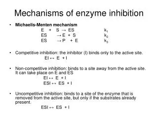
Similar Enzymes • Catalytic domain of ETA is functionally and structurally similar to both mono-ADPRTs and PARPs • Diphtheria toxin (DT) • Mono-ADPRT that also catalyzes the ADP-ribosylation of eEF2 • PARPs (Poly-(ADP-ribosyl) polymerases) • Eukaryotic nucleus • Catalyze the covalent attachment of ADP-ribose units from NAD+ to itself and nuclear DNA-binding proteins • Responds to DNA strand breakage • Rapid activation of PARP depletes NAD+ within the cell • Disruption of energy production processes
The Inhibitors ( ) n=0 or 1 • Common structural motif of inhibitors • Benzamido group fused into a heteroring to lock amide in s-trans conformation • Mimics the nicotinamide moiety of NAD+ • R-group substitutions that include the addition of hydrogen donors and/or acceptors to increase water solubility Nicotinamide moiety of NAD+
Tricyclic Lactams – [6,6,6] Ring System PJ34 GP-L GP-M GP-G GP-N
Tricyclic Lactams – [5,6,7]-Ring Systems GP-D GP-F GP-H GP-I
Other Inhibitors Classes Bicyclic Lactam Tetracyclic Lactam 5-AIQ GP-P NAD+ Analog 2’-F-ribo-NAD+
Looking at the 3D Inhibitor Structures • The Dundee PRODRG2 Server • http://davapc1.bioch.dundee.ac.uk/programs/prodrg/prodrg.html • Generates • PDB file (with and without hydrogens) • X-ray refinement topology and parameter files for use with CNS • CHECK THIS WEBSITE OUT! • Let’s look at the inhibitor structures in 3D
Correlate IC50 to Structure GP-D PJ34 GP-M IC50 = 165 nM Planar IC50 = 284 nM Planar IC50 = 287 nM Planar GP-P GP-F GP-L IC50 = 453 nM Non-Planar IC50 = 478 nM Non-Planar IC50 = 610 nM Non-Planar
Correlate IC50 to Structure GP-G GP-H GP-N IC50 = 688 nM Planar *Exception IC50 = 964 nM Non-Planar IC50 = 1.05 mM Planar *Exception GP-I 5-AIQ F-NAD+ IC50 = 4.46 mM Non-Planar IC50 = 22.8 mM Planar IC50 = 82.4 mM Planar
Summary of Inhibitors • Importance of a locked benzamido group • More potent inhibitors have a core ring structure that is planar • Exceptions are GP-G and GP-N • Piperazine moieties as their R-group • Unfavourable • Active site prefers compounds that are more rigid and compact • Non-flat ring systems may be too big to fit into the active site • Positioning of the hydrogen bonding lactam is critical • GP-D is the most potent inhibitor in this study • [5,6,7] tricyclic lactam containing a indole • Potential H-bond to Glu-553 analogous to PARPs?
PJ34 – Its History • Originally synthesized by Inotek Pharmaceuticals to target PARP • Now commercially available from Sigma • Water-soluble phenanthridinone derivative • Well-characterized compound • in vitro and in vivo studies in several PARP related systems • Stroke, heart disease and transplantation, diabetes, cancer, exposure to cytotoxic oxidants etc….
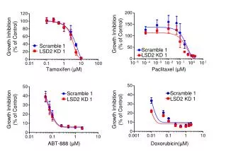
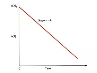
Inhibition of PE24H by PJ34 • Characterization of PJ34 • IC50 = 284 ± 69 nM • KD = 820 ± 54 nM (A) • 70x tighter binding to PE24H compared to NAD+ • Competitive inhibitor (B) • Preliminary work using wheat germ eEF2 suggest that the Ki is ~300 nM A B
Let’s Crystallize PE24H with PJ34! Make Crystals Collect Diffraction Data Molecular Replacement Build Model with ‘O’ Refinement with CNS Check Quality of Model with PROCHECK Create Figures using PyMOL
The Program ‘O’ • Macromolecular crystallographic modeling tool • used to look at macromolecular structures, analyze them, compare them, modify them and to build them from scratch • Helpful website: • http://xray.bmc.uu.se/alwyn/A-Z_of_O/everything.html
CNS • Crystallography & NMR System (CNS) • international collaborative effort among several research groups • designed to provide a flexible multi-level hierarchical approach for the most commonly used algorithms in macromolecular structure determination • include heavy atom searching, experimental phasing (including MAD and MIR), density modification, crystallographic refinement with maximum likelihood targets • Website: • http://cns.csb.yale.edu/v1.0/ • Copy scripts from the website and save them in XEmacs • Run CNS after scripts have been edited in XEmacs
PE24H-PJ34 Structure Monomer A Monomer B • Includes residues 399 to 602 • Poor density for C-terminal residues, 603 to end • Monomers A and B superimpose with only minor alterations • Monomer B • Residues 459 to 464 unresolved
PE24H-PJ34 Structure Monomer A Lys-590 Glu-522 Monomer B Crystal Packing
Interactions of PJ34 within the Active Site • Hydrophobic Pocket • 60% of the surface of PJ34 buried within the toxin • Trp-466, Tyr-470, Ile-471, Ala-472, Leu-477, Ala-478 and Tyr-481
Omit Map of PJ34 within the Active Site Gln-485 Tyr-470 Glu-553 Tyr-481 Gly-441 Leu-477 Ala-478 Ala-472 His-440 2Fo-Fc omit map of PJ34 bound within the active site of ETA contoured at 1.
PJ34-PE24H Structure 1 = 2.74 Å, 2= 2.45 Å, 3 = 2.53 Å, 4=3.08 Å Tyr-481 is 4 Å and Tyr-470 is 6-7 Å (and at 40°) away from PJ34 Glu-553 Leu-477 Ala-478 Tyr-481 Tyr-470 4 Ala-472 1 2 3 Gln-485 Gly-441 His-440
Superposition of Modeled Loop Toxin-b-TAD Li et al., 1996 Toxin-PJ34 Toxin with hydrolyzed NAD+ Li et al., 1995
Comparisons with DT and PARP Tyr-470 PJ34 Tyr-481 LOOP Ala-478 Glu-553 Ala-472 Gly-441 His-440 Tyr-470 NU1025 LOOP Tyr-481 Ala-478 Glu-553 Gly-441 Ala-472 His-440 PE24H-PJ34 vs DT PE24H-PJ34 vs PARP
Final Thoughts • First report of a structure of a mono-ADPRT in complex with an inhibitor • Confirmed the hydrogen bonding to the lactam moiety as seen in PARP and as predicted earlier with the NAP-ETA model • Planar compounds may sandwich better into the nicotinamide binding pocket than more flexible compounds • Steric interactions? • Similarities and differences between ETA/DT and PARP • Exploit the differences to preferentially target one enzyme over the other