Download
1 / 12
120 likes | 293 Vues
Evaluation by Optical Coherence Tomography (OCT) of Neointimal Coverage of Sirolimus -Eluting Stent Three Months After Implantation.

E N D
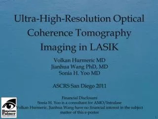
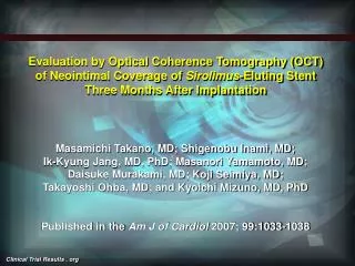
Evaluation by Optical Coherence Tomography (OCT) of Neointimal Coverage of Sirolimus-Eluting Stent Three Months After Implantation Masamichi Takano, MD; Shigenobu Inami, MD; Ik-Kyung Jang, MD, PhD; Masanori Yamamoto, MD; Daisuke Murakami, MD; Koji Seimiya, MD; Takayoshi Ohba, MD; and Kyoichi Mizuno, MD, PhD Published in the Am J of Cardiol 2007; 99:1033-1038
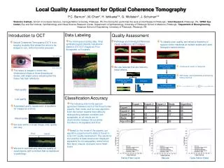
Evaluation by OCT of Neointimal Coverage of Sirolimus-Eluting Stent Three Months After Implantation: Background • The US FDA recommends that aggressive medical therapies with dual antiplatelet drugs be continued > 3 months after sirolimus-eluting stent (SES) implantation for the prevention of stent thrombotic occlusion. • Nevertheless, there is no evidence of completion of reendothelialization after SES implantation at 3 months in living patients, and late stent thrombosis has been reported in patients with SES. • Therefore, optical coherence tomographic (OCT) analysis at 3-month follow-up, focusing on stent exposure and malapposition of a SES, might provide adequate information on the safety of discontinuation of dual antiplatelet therapy for patients. Takano, et al. Am J Cardiol 2007; 99: 1033-38
Evaluation by OCT of Neointimal Coverage of Sirolimus-Eluting Stent Three Months After Implantation: Design 21 patients (21 lesions) undergoing percutaneous coronary intervention (PCI) with SESs for their native coronary arteries * Three month follow-up (90 ± 10 days) Exclusion Criteria: unprotected left main coronary artery disease, restenotic lesions, chronic renal failure without regular hemodialysis, and severely decreased left ventricular systolic function. 3 mos. after SES implantation • Evaluation of stent exposure and malapposition using OCT in different clinical presentations, such as acute coronary syndrome (ACS) and non-ACS. • Performance of motorized optical coherence tomographic pullback (1 mm/s) to examine consecutive implanted 31 SESs in 21 lesions. • Measurement of NIH thickness inside each strut and percent neointimal hyperplasia (NIH) area in each cross section. * All patients received dual antiplatelet drugs (Ticlopidine 200mg/day and aspirin 100 mg/day) Takano, et al. Am J Cardiol 2007; 99: 1033-38
Evaluation by OCT of Neointimal Coverage of Sirolimus-Eluting Stent Three Months After Implantation : Baseline Characteristics • Study subjects consisted of 9 patients with ACS (3 with STEMI, 3 with non-STEMI, and 3 with unstable angina) and 12 with non-ACS. • Patients’ characteristics were similar between the ACS and non-ACS groups. Takano, et al. Am J Cardiol 2007; 99: 1033-38
Evaluation by OCT of Neointimal Coverage of Sirolimus-Eluting Stent Three Months After Implantation : Results • In total, 4516 struts in 567-mm single-stented segments were analyzed. • Overall, NIH thickness and percent NIH area were 29 ± 41 μm and 10 ± 4%, respectively. • Rates of exposed struts and exposed struts with malapposition were 15% and 6%, respectively. • These were more frequent in patients with ACS than in those with non-ACS (18% vs. 13%, p < 0.0001; 8% vs. 5%, p <0.005, respectively). Takano, et al. Am J Cardiol 2007; 99: 1033-38
Evaluation by OCT of Neointimal Coverage of Sirolimus-Eluting Stent Three Months After Implantation A B C Figure 1. OCT measurements in length and area. (A) Typical cross-sectional image of single-stented segment. Six stent struts with shadowing are clearly recognized. Magnification of the image is displayed in the lower left. Numerals on vertical and horizontal axes indicate absolute length (millimeters). (B) Length between surfaces of the neointima and each stent strut (NIH thickness). Each NIH thickness is displayed on the upper left. (C) Stent area (outer circle) and lumen area (inner circle) were measured by manual trace. Each area measurement is displayed on the upper left. C Takano, et al. Am J Cardiol 2007; 99: 1033-38
Evaluation by OCT of Neointimal Coverage of Sirolimus-Eluting Stent Three Months After Implantation A A B C Figure 2. Different nonexposed strut types with malapposition. Although all struts were diagnosed as malapposed, there were intracoronary structures on them. (A) Struts seem to float into the lumen compared with the extra-stent lumen (arrowheads). (B) A strut is surrounded by an intracoronary structure with an irregular surface. This OCT finding likely indicates that a part of the analyzed strut has fibrin deposition. (C) Maximum distance between the strut surface and the vessel wall is 200 μm. This strut is completely buried under the intracoronary structure protruding into the lumen. Takano, et al. Am J Cardiol 2007; 99: 1033-38
Evaluation by OCT of Neointimal Coverage of Sirolimus-Eluting Stent Three Months After Implantation Figure 2 A A B C Figure 3. Different exposed strut types. (A) An exposed strut without malapposition has no structure inside the strut, which adheres to the vessel wall. (B, C) Exposed struts with malapposition with a maximum distance < 160 μm between the strut surface and the vessel wall. (C) No filling space between the struts and adjacent vessel wall was found. Takano, et al. Am J Cardiol 2007; 99: 1033-38
Evaluation by OCT of Neointimal Coverage of Sirolimus-Eluting Stent Three Months After Implantation Figure 4. Intracoronary thrombus with an obvious protruding mass between 4 and 5 o’clock. Takano, et al. Am J Cardiol 2007; 99: 1033-38
Evaluation by OCT of Neointimal Coverage of Sirolimus-Eluting Stent Three Months After Implantation : Limitations • Findings were based on observations in a relatively small number of patients and stented segments. • This study was not randomized. Lesion characteristics such as reference diameter before PCI and stent and lumen areas at follow-up differed between the two groups, and so relative NIH area was compared. Takano, et al. Am J Cardiol 2007; 99: 1033-38
Evaluation by OCT of Neointimal Coverage of Sirolimus-Eluting Stent Three Months After Implantation : Limitations Cont. • Although OCT was used, detection of very thin structures below its image resolution and certain distinctions between fibrin and NIH were impossible. • Occurrence of stent malapposition was unidentified because OCT examination was performed at a single time point. Takano, et al. Am J Cardiol 2007; 99: 1033-38
Evaluation by OCTof Neointimal Coverage of Sirolimus-Eluting Stent Three Months After Implantation: Summary • In conclusion, the present study using optical coherence tomography demonstrated the existence of malapposed struts without neointimal coverage and incomplete neointimal proliferation at 3 mos. after SES implantation. • However, it is not clear that these uncovered struts are associated with clinical events such as late stent thrombosis. • This study suggests that dual antiplatelet therapy might be continued >3 months after SES implantation. Takano, et al. Am J Cardiol 2007; 99: 1033-38