Download
1 / 22
230 likes | 305 Vues
Explore the growth and characterization of single crystal CVD diamond detectors for alpha particle and neutron detection, including structural analysis and radiation monitoring performance. Learn about the growth parameters, structural characterization, efficiency, and charge collection distance of these detectors. Discover the impact of pumping on detector performance and the behavior of diamonds when used in particle detection applications.

E N D
SINGLE CRYSTAL CVD DIAMOND DETECTORS Cristina Tuvè Department of Physics and Astronomy, University of Catania and INFN Cristina.Tuve@ct.infn.it M. Angelone1, V. Bellini4, A. Balducci2, M.G. Donato3, G. Faggio3, M. Marinelli2, G. Messina3, E. Milani2, M.E. Morgada2, M. Pillon1, R. Potenza4, G. Pucella2, G. Russo4, S. Santangelo3, M. Scoccia2, C. Sutera4, A. Tucciarone2, G. Verona‑Rinati2 1 Associazione EURATOM-ENEA sulla Fusione, Frascati (Roma), Italy 2 Dip. di Ing. Meccanica, University of Roma “Tor Vergata”, Italy 3 Dip. Meccanica dei Materiali, University of Reggio Calabria, Italy 4 Department of Physics, University of Catania ad INFN, Italy
Diamond films were grown in the Laboratories of Roma “Tor Vergata”University by CVD Polycristalline CDV diamond --particle detection - 12C particle detection at National Southern Laboratories (LNS) in Catania (Italy) - Neutron monitoring at Joint European Torus (JET) in Culham (U.K.) Single crystal CVD diamond growth Characterization Radiation detectors - -particle detection - Neutron detection Conclusions OUTLINE
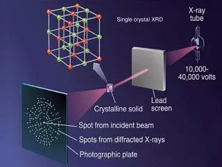
Structural characterization Diamonds are grown in the Laboratories of Roma “Tor Vergata”University by MicroWave Plasma Enhanced Chemical VaporDeposition (MWPECVD) Growth Parameters:Plasma CompositionCH4 / H2 1% gas mixture Temperature 750 ºC Growth rate 0.7 mm/h • Micro-Raman and Micro-PL • FWHM = 2.4 cm-1, comparable with that of natural mono-crystals (FWHM=2 cm-1) • PL band practically absent (APL/Ap<1/60) M.G. Donato, G. Faggio, G. Messina, S. Santangelo Dip. Meccanica e Materiali Università di Reggio Calabria - Italy
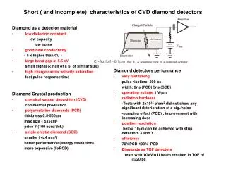
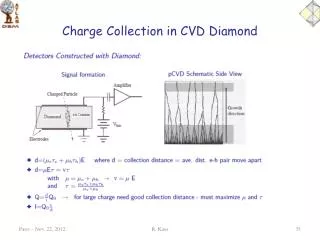
Ionizing particle Au electrode Out Charge Sensitive Au contact 7 mm2 wide 100 nm thick Ag contact paint Amplifier x e h CVD Diamond p - Si Bias Ag electrode Qc : charge induced for each e-h pair L : detector thickness e : charge of the carriers x : total distance the e and h move apart • Efficiency h = QC/Q0 • QC :collected charge • Q0 : total charge • Charge Collection Distance (CCD) : d = le + lh = (me te +mhth) E l: mean drift distance m : mobility t :lifetime E: applied electric field The response of the detector is related to the presence of traps Diamond Detectors Samples showing a very narrow Raman peak and extremely low photoluminescence background can present very different behaviour when used as particle detectors Particle detection can be used as a very sensitive probe for carrier trapping characterization
PolyCVD Diamond detector Effect of pumping Pumping is the procedure to saturate types of defects by means of -particles from 90Sr. Increased efficiency h and charge colection distance after ioniziong radiation exposure 241Am -particle spectra as measured by a policrystalline CVD diamond sample • Low energy resolution • The dispersion around mean efficiency depends of the • fluctuations of the defect density . M. Marinelli et al. Applied Physics Letters 75 (1999) 3216
Negative Polarization _ G h l Positive bias – Pumped e D + Positive Polarization + e l h _ lh >> le Because h+ > h- a-Spectra: PolyCVD diamond Negative bias – Pumped
Si detector Beam L.N.S. Tandem accelerator Gold Faraday target cup Diamond detectors Vacuum pumps Gold contact ˆ n q Incident particles Rotating sample holder Diamond sample LNS - Experimental set-up Target:Au300mg/cm2 Beams:12C, 6Li Beam Energy:12C: 16÷91 MeV We mounted the samples on a rotating holder. In order that, the penetration depth of the incident particles could vary over a factor of 6. The incidence angle q varied in the 0o ÷ 80o range. Combining both the angle and energy scans, the penetration depth can be varied from2m to 85 m.
Negative Polarization _ G h d e L Positive Polarization + + e d h _ le= 1/ae = 0.8mm; lh=1/ah=33mm • Defects are concentrated near the substrate side Nuclear particle penetration as probe of defect distribution: results E= 1 V/mm L= 55 mm pumped 12C hit the detector on the growing side in all cases
x • In grain defects: uniformly distributed insidethe sample; not dependent on position • Grain boundaries defects: concentrated at thesubstrate side; same contribution for both carrier types In-grain defects Grain boundaries defects Grain Boundaries effect model Electron and hole contributions to the conduction mechanism were successfully separated and evaluated. R.Potenza and C.Tuvè,Carbon: the future for advanced technology applications Springer series Topics in Applied Physics
Neutron detection (JET) Diamond detector installed at Joint European Torus (JET) in a tokamak (Toroidal Kamera Magnitnaya) Monitoring of the 14 MeV neutrons produced by the D-T plasma during the Trace Tritium Experiment (TTE) lasted 5 weeks CVD Diamond detector count rates Vs Silicon detector ones for all the TTE campaign at JET (5 weeks) • Very similar trend for the three detectors • Lower statistic for diamond • Smaller sensitive volume • Quite far from the plasma (about 7 m) • Good stability over 5 week of uninterrupted operation Comparison between the temporal response of CVD diamond, Silicon detector and Fission chamber for a JET pulse M. Angelone et al.,Rev.of Sc. Instr 76(2005)013506
Poly CVD diamond detectors low stability – low reproducibility • It is possible use pCVDs as counters • Dosimeters • Beam monitors • Neutron flux monitors Low Energy Resolution Why would we like to grow CVD diamond on natural diamond ? To proceed towards better energy resolution, we observed that defects concentrate near the substrate onwhich the growth begins; this can be explained with the fact that Si substrate and diamond crystals have different reticular constants, so that the first shellsin the initial growth are not free of defects and produce a columnar growth of thewhole diamond deposit.(Heteroepitaxial growth) Diamond:reticular constant: 3.561 Amg Silicon:reticular constant: 5.43086 Amg So:we attempted homoepitaxial growth using reactor parameters that optimize heteroepitaxial grown crystals.
COOLING Microwave Chemical Vapor Deposition MW-CVD Typical growth parameters Plasma composition H2-CH4, CO2-CH4 etc. Temperature 650 - 950 °C Microwave power 600 - 1000 W Pressure 50 - 130 mbar Gas flow rate 40 - 200 sccm • Substrates • (100) HPHT (Element 6, Sumitomo, others) • Natural diamond • Type Ib, IIa, B-doped
Oscilloscope Ionizing particle Shaping Amplifier Charge amplifier e h CVD diamond Multichannel analyzer Bias HPHT substrate Particle detector • CVD Layer 110 m thick • Substrate: B-doped HPHT diamond, 315 m thick • Circular Al contacts: 3mm diameter, 100nm thick
EM transverse view of a CVD diamond grown on Si substrate(columnar growth of the whole diamond deposit) EM transverse view of a CVD diamond grown on natural diamond substrate Comparison between hetero - homoepitaxially grown diamonds: transverse view
EM surface view of a CVD diamond grown on Si substrate EM surface view of a CVD diamond grown on natural diamond substrate CENTER BOUNDARY Comparison between hetero-homoepitaxially grown diamonds: surface view
X-ray diffraction and Raman spectra Micro Raman • 514 nm Ar laser • Peak @ 1332.9 cm –1 • FWHM = 1.8 cm –1 (including instrum. broadening) • Extremely low photoluminescence background • Homogeneous results all over the sample surface • Lattice parameter: d=3.567 Å M.G. Donato, G. Faggio, G. Messina, S. Santangelo Dip. Meccanica e Materiali Università di Reggio Calabria - Italy
Poly crystal • Very poor resolution • Strongly sensitive to “Priming” effect Polycrystal – Single Crystal detectors • Single Crystal Diamond • High resolution • No priming (pumping) effects
Energy resolution and peak energy vs. Voltage • The best resolution is obtained with electric fields E ≈ 1V/mm • Peak energy saturates at sufficiently high applied voltages
a-particles detection Triple source (239Pu, 241Am, 244Cm) emitting 5.16 MeV, 5.48 MeV and 5.80 MeV ‑particles respectively The detector is in the “as grown state” this means that no “priming” procedure was adopted. The same results are observed after 5 krad 90Sr particle irradiation (i.e. our usual procedure to drive our sample in a fully pumped state). • High detection efficiency • High resolution ( 1.1% ) • Low noise • No pumping effects • Good stability
Ti – T target θ = 0° The D-T fusion reactions is simulated inside the neutron transport MCNP code 0° E = 14.8 MeV, FWHM = 0.5 MeV 90° E = 14.1 MeV, FWHM = 0.3 MeV θ 260 keV Deuterium beam 241Am α-particle source Collimator θ = 90° Diamond detector Frascati Neutron Generator (FNG) • D-T fusion reactions neutrons • Neutron energy and energy-spread depends on the emission angle with respect to the D beam Neutrons and particles simultaneously irradiated
Neutron detection (FNG) Simultaneous neutrons and 241Am -particle irradiation. Calibration obtained by scaling the horizontal axe with respect to the 241Am peak • Correct position of both 0° Position measurement 90° Position measurement 12C(n,)9Be peak E = 9.1 MeV FWHM = 0.5 MeV 12C(n,)9Be peak E = 8.4 MeV FWHM = 0.3 MeV Correct position Same of the incident neutrons
The reliable use of polycrystalline diamond for neutron detection in fusion experiments has been demonstrated. High quality single crystal diamond films were homoepitaxially grown by CVD onto HPHT diamond substrates. Single crystal diamond particle detectors were produced and tested with -particle, and neutrons. A resolution of 1.1% (including electronic noise) have been measured when irradiating with -particles. No“priming” or “pumping” effect was observed. 14 MeV Neutron spectra showed a clear 12C(n, )9Be peak with a energy resolution lower than 3.6% (energy spread of the incident neutrons). CONCLUSIONS