Download
1 / 41
430 likes | 495 Vues
Explore obstructive pulmonary diseases like bronchial asthma, chronic bronchitis, and emphysema. Learn about causes, symptoms, diagnosis, and treatment options including prevention strategies.

E N D
Obstructive Pulmonary Disease Dr. Tarek Atia
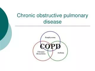
Characterized by airway obstruction that is increased with expiration. More force is required to expire a given volume of air, or emptying the lungs is slowed, or both. The main symptoms of obstructive pulmonary disease are dyspnea, and wheezing. The most common obstructive diseases are BronchialAsthma, Chronic Bronchitis, and Emphysema.
COPD Asthma Emphysema PathologicDiagnosis Physiologic Diagnosis Chronic Bronchitis Clinical Diagnosis
1- Bronchial Asthma More intermittent and acute than other types of COPD. It is a reversible disorder It occurs at all ages. Runs in families, so genetics evidence plays a role. Environmental factors interact with inherited factors to increase the risk of asthma.
Pathophysiology • Chronic Inflammatory Disorder • Results in widespread air way obstruction. • Induced by a trigger, which causes release of histamine resulting in broncho-spasm and bronchial edema. • The main pathology in acute asthma attack include: • Bronchiolar constriction, or smooth muscle spasm • Mucus hyper-secretion, or production of thick mucus • Inflammatory mucosal swelling (edema), with vascular congestion.
Clinical Manifestations Dyspnea Sometimes, cough Wheezing During remission individual is asymptomatic and pulmonary function tests are normal
Treatment • Treatment goals: • Correct hypoxia. • Reverse bronchospasm. • Reduce inflammation. • Maintain the airway. • Support breathing. • High-flow oxygen or assisted ventilations as indicated.
Special Case of Asthma • Status Asthmaticus • A severe and prolonged type of bronchial asthma that cannot be relieved by the ordinary treatment. • Great diminished breath sounds. • Recognize imminent respiratory arrest. • Aggressively manage airway and breathing. • Transport immediately.
2- Chronic Bronchitis Hyper-secretion of mucus and chronic productive cough for at least 3 months of the year for at least two consecutive years. Incidence may be increased up to 20 times in smokers and more in persons exposed to air pollution.
The main pathology in chronic bronchitis include: Inflammatory cell infiltration into the bronchial wall. Causes bronchial edema and increases size and number of mucus secreting cells. Accumulation of thick mucus (can’t be cleared because of impaired ciliary function). Increases susceptibility to infection and injury
Initially affects only larger bronchi; later on, all airways are involved. Airways collapse in early expiration, blocked by mucus leads to hypo-ventilation and Hypoxemia Air trapping prevents respiratory muscles from functioning efficiently causing barrel chest. Barrel Chest
Treatment Best treatment is PREVENTION. Stop smoking ------ progression of the disease Bronchodilators, expectorants, and chest physical therapy. Acute attacks may require antibiotics, and steroids. Oxygen therapy may be required
3- Emphysema Abnormal, permanent enlargement and destruction of the alveolar walls. Obstruction results from changes in lung tissue rather than mucus production and inflammation (pathological changes). Major mechanism is loss of elastic recoil
Pathogenesis: or causes • Exposure to irritant substances • Exposure results in the destruction of the walls of the alveoli. • Weakens the walls of the small bronchioles results in increase residual volume. • Polycythemia
The major cause is smoking Other causes are air pollution and recurrent respiratory infections Primary emphysema linked to an inherited deficiency of the enzyme alpha 1- antitrypsin which can affect lung tissue.
Emphysema • Physical Exam • Barrel chest. • Prolonged expiration and rapid rest phase. • Thin. • Pink skin due to Polycythemia. • Hypertrophy of accessory muscles.
Pathophysiology: pathological changes Begins with the destruction of the alveolar septa, which eliminates portions of the capillary bed, and increases the volume of air in the alveolus. Continued alveolar loss and loss of elastic recoil Expiration becomes difficult, causing hyper-expansion of the chest
Clinical Manifestations Dyspnea at rest Barrel chest Minimal wheezing Prolonged expiration Hypoventilation and hypoxia
Atelectasis • Collapse or incomplete expansion of part or all of the lung • Types: • Resorption: obstruction of airway. • Compressive: pleural effusion or pneumothorax.
Bronchiectasis • Dilatation of bronchi and bronchioles secondary to chronic inflammation • Associated conditions • Obstruction • Cystic fibrosis • Immotile cilia syndromes • Necrotizing pneumonia
Pulmonary Hypertension Secondary (most common): Chronic obstructive pulmonary disease Chronic interstitial pulmonary disorders Chronic heart failure Recurrent pulmonary emboli Primary (idiopathic)
Respiratory Failure (RF) The inability of the lungs to adequately oxygenate the blood and to clear it of carbon dioxide. Respiratory Failure could be :- Acute RF Chronic RF
Chronic respiratory failure • Due to progressive hypoventilation from airway obstruction or restrictive disease • Causes of Acute RF: • Pulmonary embolism • Direct injury to the lungs, airways or chest wall • Indirect due to injury of the brain. • Acute Respiratory Distress Syndrome (ARDS)
Respiratory failure always presents with:- • Dysnpeaalways present, but may be difficult to detect a change in a chronic patient • Since nervous tissue is highly oxygen-dependent • Drowsiness, memory & visual impairment, and headache are commonly seen.
Two Principal Patterns: 1- Hypoxic Respiratory Failure 2- Hypoxic-Hypercapnic Respiratory Failure
Seen when pO2 {oxygen partial pressure} falls to or below 60 mm Hg Typically seen in:- Chronic bronchitis and emphysema. Lung consolidation due to infection (pneumonia). In lung collapse, Pulmonary hypertension 1- Hypoxic Respiratory Failure:
2- Hypoxic-Hypercapnic RF When arterial pCO2 {Carbon dioxide partial pressure} (normally 40 mm Hg) exceeds 45 mm HG, condition is called hypercapnia. Caused by airway obstructive conditions, and hypoventilation from CNS problem, thoracic cage or neuromuscular abnormalities.
Evaluation and treatment Diagnosis based on physical examination, blood gases and imaging Treatment is based on early detection, supportive therapy and prevention of complications, especially infection. Often requires mechanical ventilation.
Carcinoma of the lung 7% of all deaths More common in males. • Causes • 85-95% smoking • 1% asbestos + smoking (estimate) • Rare arsenic, chromium, nickel...
Major types of lung carcinoma Small cell carcinoma 25% Non-small cell carcinoma Squamous cell carcinoma 35% Adenocarcinoma 30% Large cell carcinoma 10%
Small cell carcinoma Frequency: 25 % Smoking: 95% of patients Males >> females Survival (5 years): 1 - 5 %.
Squamous cell carcinoma Frequency: 35% Smoking: X 25 (increased risk) Males > females Survival (5 years): 15 - 20% Arises in bronchial squamous metaplasia Centrally located
Large cell carcinoma Frequency: 10 % Gross picture: Peripheral lesion Microscopic Wastebasket group of tumors that do not fit the criteria of a squamous cell carcinoma, adenocarcinoma, or small cell carcinoma Prognosis: Similar to adenocarcinoma
Adenocarcinoma Frequency: 30% Smoking: X 3 (increased risk) Males < females Survival (5 years): 15 - 20% Peripheral
Bronchiolo-Alveolar carcinoma Frequency: 2 % Smoking Males = females Survival (5 years): 25-40 %. Single or multiple tumor nodules
Mesothelioma Malignant tumor of mesothelial cells Highly malignant neoplasm with short survival Most patients (70%) have a history of asbestos exposure Asbestos exposure also increases the risk of pulmonary cancer Smoking is not related to mesothelioma