Download

1 / 19
250 likes | 631 Vues
Stages of Development of Blood Cells. Dr. Sama ul Haque. Objectives. Understand the composition of whole blood. How to prepare a blood smear. Describe the structure of Erythrocyte. Enlist different types of leucocytes. Explain the differentiation of myeloid and lymphoid stem cells.

E N D
Stages of Development of Blood Cells Dr. Sama ul Haque
Objectives • Understand the composition of whole blood. • How to prepare a blood smear. • Describe the structure of Erythrocyte. • Enlist different types of leucocytes. • Explain the differentiation of myeloid and lymphoid stem cells. • Discuss the development of blood cells in red bone marrow.
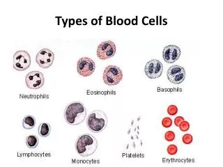
Types of Human LeucocytesNeutrophils, eosinophils, and basophils have granules that stain specifically with certain dyes and are called granulocytes. Lymphocytes and monocytes are considered agranulocytes, even though they may show azurophilic granules (lysosomes), which are also present in other leukocytes.
NeutrophilsNeutrophils can be identified by their multilobulated nuclei, with lobules held together by thin strands. With this feature the cells are often called polymorphonuclear leukocytes, or just polymorphs.
EosinophilsEosinophils are about the same size as neutrophils but have bilobed nuclei and abundant coarse cytoplasmic granules. The cytoplasm is often filled with brightly eosinophilic specific granules, but also includes some azurophilic granules. (a):An eosinophil next to a neutrophil for comparison with its nucleus and granules. (b): Even with granules filling the cytoplasm, the two nuclear lobes of eosinophils are usually clear.
BasophilsApproximately the same size as neutrophils and eosinophils, but have large, strongly basophilic specific granules which usually obstruct the appearance of the nucleus having two or three irregular lobes.
Lymphocytes Lymphocytes are agranulocytes and lack the specific granules characteristic of granulocytes. (a):Small lymphocytes are slightly larger than the neighboring erythrocytes with spherical nucleus. (b): Medium lymphocytes are distinctly larger than erythrocytes. (c): Large lymphocytes, much larger than erythrocytes.
MonocytesMonocytes are large agranulocytes that circulate as precursors to macrophages and other cells of the mononuclear phagocyte system. Monocytes show their eccentric nuclei indented, kidney shaped, or U shaped.
PlateletsPlatelets are cell fragments 2–4 µm in diameter derived from megakaryocytes of bone marrow. Their primary function is to rapidly release the content of their granules upon contact with collagen to begin the process of clot formation and reduce blood loss from the vasculature. Platelets (arrows) are often found as aggregates.
Differentiation of Myeloid And Lymphoid Stem Cells 1. EPO– Erythropoietin 2. CSF– Colony Stimulating Factor 3. GM-CSF– Granulocyte+Macrophage CSF 4. M-CSF– Macrophage CSF 5. G-CSF– Granulocyte CSF