Chapter 1
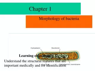
Chapter 1. Morphology of bacteria. Learning objectives: Understand the structural features that are important medically and for identification. KEY WORDS. Cell envelope Lipopolysaccharide 脂多糖 (endotoxin 内毒素 ) Cell wall Teichoic acid 磷壁酸

Chapter 1
E N D
Presentation Transcript
Chapter 1 Morphology of bacteria Learning objectives: Understand the structural features that are important medically and for identification
KEY WORDS Cell envelope Lipopolysaccharide脂多糖(endotoxin内毒素) Cell wall Teichoic acid 磷壁酸 Cell membrane Teichuronic acid Outer membrane Lipoteichoic acid 脂磷壁酸 Braun lipoprotein Undecaprenol (bactoprenol) Endospore 芽孢 Porins细胞外膜孔道蛋白
Key Words Periplasmic space周浆间隙 Spheroplast/protoplast原生质体 Chromosome 染色体 Flagella鞭毛 Permeases 透明质酸酶 lipoprotein脂蛋白 Plasmid质粒 Axial filament轴丝 Peptidoglycan肽聚糖 (murein, mucopeptide粘肽) Periplasmic binding protein Storage Granules Gram stain Pili,菌毛(fimbriae) Gram negative Capsule荚膜 (slime layer, glycocalyx) Gram positive Ribosome核糖体
I. Size • Unit for measurement:Micron or micrometer,μm: 1μm=10-3mm • Size: Varies with kinds of bacteria, and also related to their age and external environment.
Sizes • The unit of measurement • micrometer (μm)
II. Basic shapes • Spherical: coccus • Straight: bacillus • Curved or spiral: spiral bacterium
Shape of Bacteria • Cocci: sphere, 1μm • Bacilli: rods , 0.5-1 μm in width -3 μm in length • Spiral bacteria: 1~3 μm in length and 0.3-0.6 μm in width
Arrangements • Coccus • Diplococcus: • in pairs • Streptococcus: • in chains • Tetrad • Sarcina • Staphylococcus : • in bunches like grapes
Diplococcus Streptococcus pneumoniae
Tetrad Sarcina
III. Bacterial structures • Basic • Cell wall • Cytoplasmic membrane • Cytoplasm • nucleoid • Special • Capsule • Flagellum • Pilus • Spore
1884: Christian Gram:First publication for the Gram stain method Nearly all germs be separated violet(positive) or red (negative)by Gram.
Compositions of cell wall of G+ germ L-丙氨酸 D-谷氨酸 L-赖氨酸 D-丙氨酸 • 2.Peptidoglycan in Gram+ bacteria • Carbohydrate Backbone • Tetrapeptide side chains • pentapeptide cross bridges • Protection • Drug sensitivity • Penicillin • Lysozyme lysozyme penicillin
Peptidoglycan structure in Gram- bacteria G • Much thiner • (1-2 layers) • No cross bridges • On tetrapetide side • chain third amino • acid is Diaminopimelic • acid(二氨基庚二酸DAP) G M M 丙 谷 丙 DAP 谷 G G 丙 DAP E.coli 丙
Structures and compositions of cell wall • 25 repeating units of three to five sugars. • Outer membrane • Only found in G- bacteria • Compositions: Lipoprotein, Lipid bilayer, and LPS • five sugars linked through ketodeoxyoctulonate (KDO酮脱氧辛酸酯) to lipid A
Figure 2 Schematic structures of (A) the envelope of GNB and (B) isolated and membrane-associated LPSs and LOSs Biochemical Society Transactions www.biochemsoctrans.org Biochem. Soc. Trans. (2003) 31, 785-790
半乳聚糖 LPSR 庚糖 Schematic representation of the E.coli envelope adapted from Raetz and Withfield with minor modifications (Raetz and Whitfield, 2002). Abbreviations: LPS, lipopolysaccharide; MDO, membrane-derived oligosaccharides; Kdo, 3-deoxy-d-manno-oct-2-ulosonic acid; PPEtn, phosphoethanolamine磷酸乙醇胺
Schematic structures of the envelope of GNB • membrane-associated LPSs and LOSs • KDO, keto酮类的-deoxyo脱氧ctanoate辛酸盐/酯 ; • MDO, membrane-derived衍生的 oligosaccharides低聚糖.
B.special components ofcell wall Teichoicacid of gram positive B Only found in G+ bacteria • Two types • Wall teichoic acid • Lipoteichoic acid Role Antigen specificity Pathogenicity
B. Functions of cell wall • Gives the bacterial shape and prevents osmotic lysis • Transports materials inside and outside of bacterium • Possesses antigenic specificity • Provides many pathogenic properties
A comparison of the structure of germ-positive/negative cell wall Thick layer
pattern of peptidoglycan Negative germ positive germ
Germ is stained violet or red Step 1.Crystal violet 2.Gram’s iodine 3.Alcohol 4.safranin
Structures and compositions of cell wall After you stained the bacteria, you can seen two kinds germs Gram-positive bacteria Gram-negative Under microscopy we also seen their the shape, size arranging etc .
L forms • (As first found in the Lister laboratory) • Definition • bacteria that their peptidoglycan is destroyed or lost by with lysozyme or penicillin but they can survive under highly osmotic environment. • Two types • Protoplasts: L forms derived from G+ bacteria. Enclosed by cytoplasmic membrane. • Spheroplasts:L forms derived from G- bacteria. Enclosed by partial cell wall.
Bacteria L form • Defective cell wall-bacterial : protoplast, spheroplast
Pathogenesis of L form • Similar to the infection of virus or mycoplasma ( the organisms without wall)
culture • L forms are difficult to cultivate。They require a special high osmotic media。 Colonies : like fried egg __oval, middle thick, edge thinner. • Some L form can revert to 回复the normal bacillary form。
Colonies : fried egg Feature Colony of L form oval, middle thick, edge thiner.
All bacteria have a cell membrane where oxidative phosphorylation occurs (since there are no mitochondria). Cell Wall Cytoplasm Cell membrane Outside the cell membrane is the cell wall which is rigid and protects the cell from osmotic lysis.
Function of Cell membrane • Selective permeability and transport of solutes into cells • Electron transport and oxidative phosphorylation • Excretion of hydrolytic exoenzymes • Site of biosynthesis of DNA, cell wall polymers and membrane lipids.
Mesosomes • Definition The irregular invagination内陷 of bacterial cell membrane that is more prominent in gram-positive than in gram-negative bacteria. • Mesosomes are specialized structures formed by cytoplasmic membrane. invagination
mesosome • Its function is not definitely known, it appears to participate in DNA replication and cell division. • It also provide energy because increases surface area.
The bacterial cytoplasm is densely packed with 70S ribosomes.
Ribosomes • Ribosomes have a sedimentation coefficient(沉降系数)of 70S • That composed of 30S and 50S subunits。This is unlike the eukaryotic 80 S (40 S+ 60 S) ribosome. • They are the sites of action of many antibiotics that inhibit protein biosynthesis。