Download
1 / 36
370 likes | 642 Vues
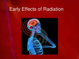
Late Effects of Radiation. EARLY EFFECTS of radiation exposure are produced by high radiation doses. Late effects of radiation exposure are the result of low doses delivered over a long period.

E N D
EARLY EFFECTS of radiation exposure are produced by high radiation doses. Late effects of radiation exposure are the result of low doses delivered over a long period. Radiation exposures experienced by personnel in diagnostic imaging are low dose and low linear energy transfer (LET). In addition, the exposures in diagnostic imaging are delivered intermittently over long periods. The principal late effects of low-dose radiation over long periods consist of radiation-induced malignancy and genetic effects. Life span shortening and effects on local tissues also have been reported as late effects, but these are not considered significant. Radiation protection guides are based on suspected or observed late effects of radiation and on an assumed linear, nonthreshold dose-response relationship.
Most late effects are also known as stochastic effects. Stochastic effects of radiation exposure exhibit an increasing incidence of response—not severity—with increasing dose. No dose threshold has been established for a stochastic response
Our radiation protection guides are based on the late effects of radiation and on linear, nonthreshold dose-response relationships.
Studies of large numbers of people exposed to a toxic substance require considerable statistical analyses. Such studies, called epidemiologic studies, are required when the number of persons affected is small. Epidemiologic studies of people exposed to radiation are difficult because: (1) the dose usually is not known but is presumed to be low, (2) the frequency of response is very low. Consequently, the results of radiation epidemiologic studies do not convey the statistical accuracy associated with observations of early radiation effects.
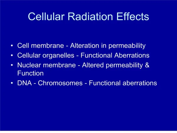
LOCAL TISSUE EFFECTS Skin In addition to the early effects of erythema and desquamation and late-developing carcinoma, chronic irradiation of the skin can result in severe nonmalignant changes. Early radiologists who performed fluoroscopic examinations without protective gloves developed a very callused, discolored, and weathered appearance to the skin of their hands and forearms. In addition, the skin would be very tight and brittle and sometimes would severely crack or flake. This late effect was observed many years ago in radiologists and is called radiodermatitis. The dose necessary to produce such an effect is very high. No such effects occur in the current practice of radiology
Chromosomes Irradiation of blood-forming organs can produce hematologic depression as an early response or leukemia as a late response. Chromosome damage in the circulating lymphocytes can be produced as both an early and a late response. The types and frequency of chromosome aberrations have been described previously; however, even a low dose of radiation can produce chromosome aberrations that may not be apparent until many years after radiation exposure. For example, individuals irradiated accidentally with rather high radiation doses continue to show chromosome abnormalities in their peripheral lymphocytes for as long as 20 years. This late effect presumably occurs because of radiation damage to the lymphocytic stem cells. These cells may not be stimulated into replication and maturation for many years.
Cataracts In 1932, E.O. Lawrence of the University of California developed the first cyclotron, a 5-inch-diameter device capable of accelerating charged particles to very high energies. These charged particles are used as “bullets” that are shot at the nuclei of target atoms in the study of nuclear structure. By 1940, every university physics department of any worth had built its own cyclotron and was engaged in what has become high-energy physics. The modern cyclotron is used principally to produce radionuclides for use in nuclear medicine especially fluorine-18 for positron emission tomography (PET). The largest particle accelerators in the world are located at Argonne National Laboratory in the United States and at CERN in Switzerland. These accelerators are used to discover the ultimate fine structure of matter and to describe exactly what happened at the moment of creation of the universe Early cyclotrons were located in one room and a beam of high-energy particles was extracted through a tube and steered and focused by electromagnets onto the target material in the adjacent room. At that time, sophisticated electronic equipment was not available for controlling this high-energy beam. Cyclotron physicists used a tool of the radiologic technologist, the radiographic intensifying screen, to aid them in locating the high-energy beam. Unfortunately, in so doing, these physicists received high radiation doses to the lens of the eye because they had to look directly into the beam.
In 1949, the first paper reporting cataracts in cyclotron physicists appeared. By 1960, several hundred such cases of radiation-induced cataracts had been reported. This was particularly tragic because there were few high-energy physicists.
Radiation-induced cataracts occur on the posterior pole of the lens.
On the basis of these observations and animal experimentation, several conclusions can be drawn regarding radiation-induced cataracts. The radiosensitivity of the lens of the eye is age dependent. As the age of the individual increases, the radiation effect becomes greater and the latent period becomes shorter. Latent periods ranging from 5 to 30 years have been observed in humans, and the average latent period is approximately 15 years. High-LET radiation, such as neutron and proton radiation, has a high relative biologic effectiveness (RBE) for the production of cataracts.
The dose-response relationship for radiation-induced cataracts is nonlinear, threshold.
If the lens dose is high enough, in excess of approximately 1000 rad (10 Gyt), cataracts develop in nearly 100% of those who are irradiated. The precise level of the threshold dose is difficult to assess. Most investigators would suggest that the threshold after an acute x-ray exposure is approximately 200 rad (2 Gyt). The threshold after fractionated exposure, such as that received in radiology, is probably in excess of 1000 rad (10 Gyt). Occupational exposures to the lens of the eye are too low to require protective lens shields for radiologic technologists. It is nearly impossible for a medical radiation worker to reach the threshold dose. Radiation administered to patients who are undergoing head and neck examination by fluoroscopy or computed tomography can be significant. In computed tomography, the lens dose can be 5 rad (50 mGyt) per slice. In this situation, however, usually no more than one or two slices intersect the lens. In either case, protective lens shields are not normally required. However, in computed tomography, it is common to modify the examination to reduce the dose to the eyes
LIFE-SPAN SHORTENING Many experiments have been conducted with animals after both acute and chronic exposures that show that irradiated animals die young. Figure below, which has been redrawn from several such representative experiments, shows that the relationship between life span shortening and dose is apparently linear, nonthreshold. When all animal data are considered collectively, it is difficult to attempt a meaningful extrapolation to humans
At worst, humans can expect a reduced life span of approximately 10 days for every rad.
Radiation-induced life span shortening is nonspecific, that is, no characteristic diseases are associated with it, and it does not include late malignant effects. It occurs simply as accelerated premature aging and death. One investigator has evaluated the death records of radiologic technologists who operated field x-ray equipment during World War II. These imaging systems were poorly designed and inadequately shielded, so that technologists received higher-than-normal exposures. Seven thousand such technologists have been studied, and no radiation effects have been observed
Risk of Life Span Shortening as a Consequence of Occupation, Disease, or Various Other Conditions
Observations on human populations have not been totally convincing. No life span shortening has been observed among atomic bomb survivors, although some received rather substantial radiation doses. Life span shortening in radium watch-dial painters, x-ray patients, and other human radiation-exposed populations has not been reported.
American radiologists have been fairly extensively studied, and early radiologists appeared to have a reduced life span. Such research has many shortcomings, not the least of which is its retrospective nature. Figure below shows the results obtained when the age at death for radiologists was compared with the age at death for the general population. Radiologists dying in the early 1930s were approximately 5 years younger than members of the general population who died at an average age. However, this difference in age at death had shrunk to zero by 1965.
The theory of radiation hormesis suggests that very low radiation doses are beneficial. Some evidence supports the principle of radiation hormesis. Radiation hormesis suggests that low levels of radiation—less than approximately 10 rad (100 mGyt)—are good for you! Such low doses may provide a protective effect by stimulating molecular repair and immunologic response mechanisms. Nevertheless, radiation hormesis remains a theory at this time, and until it has been proved, we will continue to practice ALARA—as low as reasonably achievable.
RADIATION-INDUCED MALIGNANCY All the late effects, including radiation-induced malignancy, have been observed in experimental animals, and on the basis of these animal experiments, dose-response relationships have been developed. At the human level, these late effects have been observed, but often, data are insufficient to allow precise identification of the dose-response relationship. Consequently, some of the conclusions drawn regarding human responses are based in part on animal data.
Leukemia When one considers radiation-induced leukemia in laboratory animals, there is no question that this response is real and that the incidence increases with increasing radiation dose. The form of the dose-response relationship is linear and nonthreshold. A number of human population groups have exhibited an elevated incidence of leukemia after radiation exposure—atomic bomb survivors, American radiologists, radiotherapy patients, and children irradiated in utero, to name a few.
Radiation-induced leukemia follows a linear, nonthreshold dose-response relationship.
Radiation-induced leukemia is considered to have a latent period of 4 to 7 years and an at-risk period of approximately 20 years. The at-risk period is that time after irradiation during which one might expect the radiation effect to occur. The at-risk period for radiation-induced cancer is lifetime. Data from atomic bomb survivors show without a doubt that radiation exposure to those survivors caused the later development of leukemia.
Radiologists By the second decade of radiology, reports of pernicious anemia and leukemia in radiologists began to appear. In the early 1940s, several investigators reviewed the incidence of leukemia in American radiologists and found it alarmingly high. These early radiologists functioned without the benefit of modern radiation protection devices and procedures, and many served as both radiation oncologists and diagnostic radiologists. It has been estimated that some of these early radiologists received doses exceeding 100 rad/yr (1 Gyt/yr). Currently, American radiologists do not exhibit an elevated incidence of leukemia compared with other physician specialists.
In the 1940s and 1950s, particularly in Great Britain, it was common practice to treat patients with ankylosing spondylitis with radiation. Ankylosing spondylitis is an arthritis-like condition of the vertebral column. Patients cannot walk upright or move except with great difficulty. For relief, they would be given fairly high doses of radiation to the spinal column, and the treatment was quite successful. Patients who previously had been hunched over were able to stand erect. Radiation therapy was a permanent cure and remained the treatment of choice for approximately 20 years, until it was discovered that some who had been cured by radiation were dying from leukemia. During the period from 1935 to 1955, 14,554 male patients were treated at 81 different radiation therapy centers in Great Britain. Review of treatment records showed that the dose to the bone marrow of the spinal column ranged from 100 to 4000 rad (1 to 40 Gy). Fifty-two cases of leukemia occurred in this population. When this incidence of leukemia is compared with that of the general population, the relative risk is 10:1.
Thyroid Cancer Thyroid cancer has been shown to develop in three groups of patients whose thyroid glands were irradiated in childhood. The first two groups, called the Ann Arbor series and the Rochester series, consisted of individuals who, in the 1940s and early 1950s, were treated shortly after birth for thymic enlargement. The thymus is a gland lying just below the thyroid gland that can enlarge shortly after birth in response to infection. At these facilities, radiation was often the treatment of choice. After a dose of up to 500 rad (5 Gyt), the thymus gland would shrink so that all enlargement disappeared. No additional problems were evident until up to 20 years later, when thyroid nodules and thyroid cancer began to develop in some of these patients.
Another group included 21 children who were natives of the Rongelap Atoll in 1954; they were subjected to high levels of fallout during a hydrogen bomb test. The winds shifted during the test, carrying the fallout over an adjacent inhabited island rather than one that had been evacuated. These children received radiation doses to the thyroid gland from both external exposure and internal ingestion of approximately 1200 rad (12 Gyt).
Data are just now becoming available on the nearly 100,000 persons exposed to radiation from the 1989 Chernobyl incident. No excess leukemia or cancer has been observed in this population, although a small increase in thyroid nodularity has been noted. ?
Bone Cancer Two population groups have contributed an enormous quantity of data showing that radiation can cause bone cancer. The first group consists of radium watch-dial painters. In the 1920s and 1930s, various small laboratories hired employees, most often female, who worked at benches painting watch dials with paint laden with radium sulfate. To prepare a fine point on the paintbrushes, the employees would touch the tip of the brush to the tongue. In this manner, substantial quantities of radium were ingested.
Radium salts were used because the emitted radiation, principally alpha and beta particles, would continuously excite the luminous compounds so the watch dial would glow in the dark. Current technology uses harmlessly low levels of tritium (3H) and promethium (147Pm) for this purpose. When ingested, the radium would behave metabolically similar to calcium and deposit in bone. Because of radium's long half-life (1620 years) and alpha emission, these employees received radiation doses to bone of up to 50,000 rad (500 Gyt). Seventy-two bone cancers in approximately 800 persons have been observed during a follow-up period in excess of 50 years Another population in whom excess bone cancer developed consisted of patients treated with radium salts for a variety of diseases, from arthritis to tuberculosis. Such treatments were common practice in many parts of the world until about 1950.
Skin Cancer Skin cancer usually begins with the development of a radiodermatitis. Significant data have been developed from several reports of skin cancer induced in radiation therapy recipients treated with orthovoltage (200 to 300 kVp) or superficial x-rays (50 to 150 kVp). Radiation-induced skin cancer follows a threshold dose-response relationship.
Breast Cancer Controversy is ongoing regarding the risk of radiation-induced breast cancer, with implications for breast cancer detection by x-ray mammography. Concern over such risk first surfaced in the mid-1960s, after reports were published of breast cancer developing in patients with tuberculosis. Tuberculosis was for many years treated by isolation in a sanitarium. During the patient's stay, one mode of therapy was to induce a pneumothorax in the affected lung; this was done under non–image-intensified fluoroscopy. Many patients received multiple treatments and up to several hundred fluoroscopic examinations. Precise dose determinations are not possible, but levels of several hundred rad would have been common. In some of these patient populations, the relative risk for radiation-induced breast cancer was shown to be as high as 10:1.
Lung Cancer Early in the 20th century, it was observed that approximately 50% of workers in the Bohemian pitchblende mines of Germany died of lung cancer. Lung cancer incidence in the general population was negligible by comparison. The dusty mine environment was considered to be the cause of this lung cancer. Now it is known that radiation exposure from radon in the mines contributed to the incidence of lung cancer in these miners. Observations of American uranium miners active in the Colorado plateau in the 1950s and 1960s have also shown elevated levels of lung cancer. The peak of this activity occurred in the early 1960s, when approximately 5000 miners were active in nearly 500 underground mines and 150 open-pit mines. Most of the mines were worked by fewer than 10 men; therefore, for such a small operation, one could expect a lack of proper ventilation. The radiation exposure in these mines occurred because of the high concentration of uranium ore. Uranium, which is radioactive with a very long half-life of 109 years, decays through a series of radioactive nuclides by successive alpha and beta emissions, each accompanied by gamma radiation. To date, more than 4000 uranium miners have been observed, and they have received estimated doses to lung tissue as high as 3000 rad (30 Gyt); on this basis, the relative risk was approximately 8:1. It is interesting to note that smoking uranium miners have a relative risk of approximately 20:1.
Liver Cancer Thorium dioxide (ThO2) in a colloidal suspension known as Thorotrast was widely used in diagnostic radiology between 1925 and 1945 as a contrast agent for angiography. Thorotrast was approximately 25% ThO2 by weight, and it contained several radioactive isotopes of thorium and its decay products. Radiation that was emitted produced a dose in the ratio of approximately 100:10:1 of alpha, beta, and gamma radiation, respectively. The use of Thorotrast has been shown to be responsible for several types of carcinoma after a latent period of approximately 15 to 20 years. After extravascular injection, it is carcinogenic at the site of the injection. After intravascular injection, ThO2 particles are deposited in phagocytic cells of the reticuloendothelial system and are concentrated in the liver and spleen. Its half-life and high alpha radiation dose have resulted in many cases of cancer in these organs.