Download
1 / 3
30 likes | 57 Vues
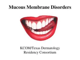
Mucous membrane pemphigoid (MMP) has been known by different names including cicatricial pemphigoid (CP), benign mucous membrane pemphigoid, benign mucosal pemphigoid, ocular pemphigus, and scarring pemphigoid.

E N D
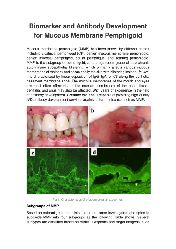
Biomarker and Antibody Development for Mucous Membrane Pemphigoid Mucous membrane pemphigoid (MMP) has been known by different names including cicatricial pemphigoid (CP), benign mucous membrane pemphigoid, benign mucosal pemphigoid, ocular pemphigus, and scarring pemphigoid. MMP is the subgroup of pemphigoid, a heterogeneous group of rare chronic autoimmune subepithelial blistering, which primarily affects various mucous membranes of the body and occasionally the skin with blistering lesions. In vivo, it is characterized by linear deposition of IgG, IgA, or C3 along the epithelial basement membrane zone. The mucous membranes of the mouth and eyes are most often affected and the mucous membranes of the nose, throat, genitalia, and anus may also be affected. With years of experience in the field of antibody development, Creative Biolabs is capable of providing high-quality IVD antibody development services against different disease such as MMP. Fig 1. Characteristics of oligodendroglial exosomes. Subgroups of MMP Based on autoantigens and clinical features, some investigators attempted to subdivide MMP into four subgroups as the following Table shows. Several subtypes are classified based on clinical symptoms and target antigens, such
as pure ocular involvement, pure oral involvement, mucosal and skin involvement, and multiple mucosal involvement. Autoantibodies are directed against various structural proteins in the epidermal basement membrane zone (EBMZ), with the 180-kD antigen (BP180) α6β4 as the main target antigen. The symptoms of MMP vary among affected individuals based on the specific site(s) involved and the progression of the disease. Blistering lesions eventually heal, sometimes with scarring. Progressive scarring may potentially lead to serious complications affecting the eyes and throat. In some cases, blistering lesions also form on the skin, especially in the head and neck area. Current researches still can’t illuminate the exact cause of MMP. Table 1. Features of MMP subgroups. Subgroup Subgroup Autoantigens Autoantigens C Clinical linical features features Pure ocular Pure ocular involvement involvement Integrin β4 subunit High-risk Pure oral Pure oral involvement involvement Integrin α6 subunit Low-risk Mucosal and Mucosal and skin skin involvement involvement BP180 Heterogeneous outcome Multiple Multiple mucosal mucosal involvement involvement Heterogenous autoantigens Heterogeneous outcome Diagnosis of MMP Usually, by using routine histopathology diagnosis, MMP can be differentiated from other mucocutaneous diseases, such as lichen planus, erythema multiforme and pemphigus vulgaris. However, it’s hard to differentiate MMP from other subepithelial autoimmune diseases depends upon neither routine histopathogy nor immunopathology. Diagnosis of MMP is often delayed by the non-specific presentations in the early stage or inconclusive biopsies. To solve the diagnosis dilemmas, the differential diagnosis must be made on the basis of combination of clinical and histopathological features. MMP is diagnosed based on a thorough clinical evaluation, a detailed patient history, identification of characteristic findings and certain tests known as a biopsy and direct immunofluorescence. For a biopsy, a small sample of skin tissue is removed
(biopsy) and microscopically examined. For direct immunofluorescence, a second biopsied skin sample is tested to detect the presence of the specific autoantibodies (e.g., IgA, IgG, and C3) that cause pemphigoid. Creative Biolabs is a well-recognized leader in the field of the custom IVD antibody discovery and development. We are dedicated to the development of innovative IVD antibodies that has higher specificity and higher sensitivity, to help increase the accuracy of the clinical diagnosis and to support clinicians in the effective management of different types of MMP. Please contact us for more information if you are interested in our services. Biomarker available now for Mucous membrane pemphigoid (MMP): BP180-NC16a Related Services: IVD Antibodies for CD164 Marker Biomarker and Antibody Development for Rheumatoid Arthritis Biomarker and Antibody Development for Childhood Asthma Biomarker and Antibody Development for SLE