Venous Return in Cardiovascular Physiology
Explore the functions of veins as blood reservoirs, central venous pressure measurement, factors influencing venous return, and more! Learn about venous incompetence, varicose veins, and the importance of venous return. Study smart for a deeper understanding.

Venous Return in Cardiovascular Physiology
E N D
Presentation Transcript
Red: very important. Green: Doctor’s notes. Pink: formulas. Yellow: numbers. Gray: notes and explanation. Venous Return Physiology Team 436 – Cardiovascular Block Lecture 7 Lecture: If work is intended for initial studying. Review: If work is intended for revision.
Objectives Study Smart: focus on mutual topics. • Discuss functions of the veins as blood reservoirs. • Describe measurement of central venous pressure (CVP) and state its physiological and clinical significance. • State determinants of venous return and explain how they influence venous return. • Define mean systemic filling pressure, give its normal value and describe the factors which affect it. • Explain the effect of gravity on venous pressure and explain pathophysiology of varicose veins. • Describe vascular and cardiac function curves under physiological and pathophysiological conditions. • Define venous return and identity the factors controlling it. • Recognize the distribution of blood in different vessels. • Explain the venous curve and relation between the venous return and the right atrial pressure. • Identify the jugular venous pressure. • Know the method of examination of the internal venous pressure.
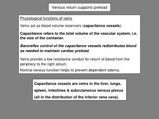
ONLY IN MALES’ SLIDES Major Component of Cardiovascular System Veins are thin-walled vessels with relatively large lumen*. • They can accommodate large changes in blood volume with little change in pressure until a limit is reached. • Beyond the limit, any change in volume results in change in pressure. Structure of veins: • All 3 layers are present**, but thinner than in arteries of corresponding size (external diameter). • Veins have paired semilunar, bicuspid valves to restrict backflow in lower extremities:- • In varicose*** veins, blood pools in veins because the valves fail causing venous walls to expand. Functions of veins • Veins are the capacitance vessels of the circulation. • Veins function as variable reservoir of blood with ≈ 2/3 blood volume contained in them. • They collect and return blood to the heart. Thus, they are important in venous return to the heart. Venous Incompetence: Skeletal muscle pump is ineffective when the valves are incompetent. • Chronically raised pressure in the veins leads to pathological distension of veins(varicose veins). • Increased capillary filtration leads to swelling (edema)with trophic skin changes and ulceration (venous ulcers). Video of (How Varicose Veins Form) Duration: (2:13)mins Lumen*: The inside space of a tubular structure, such as an artery or vein. - **3 Layers of veins and arteries: tunica intima, tunica media, tunica adventitia. Varicose veins***: Swollen and enlarged veins – usually blue or dark purple due to valve failure.
ONLY IN FEMALES’ SLIDES Veins • Veins: • Hold most of the body’s blood (70% of blood is on the venous side as they are thin-walled)& are thus called capacitance vessels. • Have thin walls & stretch easily to accommodate more blood without increased pressure (higher compliance). • Have only 0 - 10 mm Hg Pressure*. • Surrounded by skeletal muscles. *In veins: Lowest pressure = 0 - Highest pressure = 10 (Pressure rises as we move towards the arteries e.g. pressure in veins = 0 –minimum pressure-, pressure in venules = 10 –maximum pressure-)
ONLY IN MALES’ SLIDES Veins Serve as Blood Reservoirs • Normally all the blood is circulating all the time. • When the body is at rest and many of the capillaries are closed, the capacity of the venous reservoir is increased as extra blood bypasses the capillaries and enters the veins. • When this extra volume of blood stretches the veins, the blood moves forward through the veins more slowly because the total cross sectional area of the veins has increased as a result of the stretching. • Therefore, blood spends more time in the veins. • As a result of this slower transit time through the veins, the veins are essentially storing the extra volume of blood because it is not moving forward as quickly to the heart to be pumped out again. • When the stored blood is needed, such as during exercise, extrinsic factors reduce the capacity of the venous reservoir and drive the extra blood from the veins to the heart so that it can be pumped to the tissues. Remember: ARTERIES (LOW COMPLIANCE)
ONLY IN MALES’ SLIDES Venous Return • Venous return (VR) is the flow of blood back to the heart. • Under steady-state conditions, venous return (VR) must equalcardiac output (CO) when averaged over time because the cardiovascular system is essentially a closed loop. Otherwise, blood would accumulate in either the systemic or pulmonary circulations. • Venous return is determined by the difference in pressure between the venous pressure nearest to the tissues (mean systemic filling pressure; mean circulatory pressure; MCP) and the venous pressure nearest to the heart (CVP). VR CO
ONLY IN FEMALES’ SLIDES Venous Return Factors controlling venous return (Determinants of Venous Return): 1- Skeletal muscle pump → ↑ venous return. 2- Pressure drop during inspiration → ↑ venous return. Forceful expiration (Valsalva maneuver: forceful expiration against closed epiglottis; breathe and close mouth and nose and try to exhale this generates positive pressure which decreases venous return)→ ↓ venous return. 3- ↑Blood volume → ↑ venous return. 4- ↑Pressure gradient → ↑ venous return. There must be pressure difference so the blood can return to the heart. 5- ↑Venous pressure → ↑ venous return. 6- Gravity → ↓ venous return. Venous return is the quantity of blood flowing from large veins into the right atrium each min. (And it will affect the cardiac output.) (This will be explained in further detail from slide 15 to slide 21) Pressure negativity in chest cavity increases venous return, an increase in pressure difference or gradient increases venous return. Gravity decreases venous return, gravity pulls the blood in the opposite direction so it decreases venous return.
ONLY IN FEMALES’ SLIDES Venous Return Venous Return Depends On:- 1- Blood volume & venous pressure. 2- Venoconstriction caused by sympathetic NS. 3- Skeletal muscle pumps. 4- Pressure drop during inhalation. When you breathe -> increase of negative pressure in chest -> increase in venous return. Urine volume and loss of fluids increases -> decreases volume -> decreases venous return. This is the same as the previous slide but with relations between them. Very low effect, very low constriction.
ONLY IN MALES’ SLIDES Determinants of Venous Return
ONLY IN MALES’ SLIDES Cont. 2. Venous Capacity It is the volume of the blood that the veins can accommodate • It depends on the distensibility of the vein walls and the influence of any externally applied pressure squeezing inward on the veins. • At a constant blood volume, as the venous capacity → more blood spends a longer time in the veins instead of being returned to the heart → ↓ the effective circulating volume → ↓ VR. • At a constant blood volume, as the venous capacity → the MCP ↓ → ↓ VR. • As the venous capacity ↓ → VR. 1. Blood Volume • At constant venous capacity, as the blood volume → the MCP → VR. • At constant venous capacity, as the blood volume↓ → the MCP ↓ → ↓ VR. • Factors increasing blood volume: • -Decreased urine volume (water retention). • -Oral or IV hydration. • Factors decreasing blood volume: • -Dehydration. • -Diuretics. • -Increased urine volume. • -Increase in tissue fluid volume in pathological conditions e.g. Edema.
ONLY IN MALES’ SLIDES Cont. • 4. Skeletal Muscle Activity • Skeletal muscle contraction → external venous compression → ↓ venous capacity → VR (This is known as skeletal muscle pump). • Skeletal muscle activity also counter the effects of gravity on the venous system. 3. Sympathetic Activity • Venous smooth muscle is profusely supplied with sympathetic nerve fibers. • Sympathetic stimulation → venous vasoconstriction → modest in mean systemic filling pressure (MCP) → VR. • Sympathetic stimulation → ↓ venous capacity → VR. What is the effect of venoconstriction on the resistance to flow? The veins normally have such a large diameter that the moderate vasoconstriction accompanying sympathetic stimulation has little effect on resistance to flow. 5. Respiratory Activity • During inspiration there will be a decrease of pressure inside the thorax, it will be negative. Which will pull up the blood back to the heart. Thus increasing the venous return. • Valsalva maneuver is forceful expiration against a closed epiglottis and nostrils, it will make the pressure positive inside the chest, which will decrease the venous return..->
ONLY IN MALES’ SLIDES Cont. 5. Respiratory Activity (Respiratory Pump: Thoracic Pump) • As the venous system returns blood to the heart from the lower regions of the body , it travels through the chest cavity . The pressure in the chest cavity is 5 mm Hg less than atmospheric pressure. • The venous system in the limbs and abdomen is subjected to normal atmospheric pressure. • Thus , an externally applied pressure gradient exists between the lower veins and the chest veins , promoting venous return. What is the effect of Valsalva maneuver on venous return ? Return of systemic blood to the heart is impeded by the pressure inside the chest. The output of the heart is reduced and stroke volume falls. 6. Venous Valves • These valves permit blood to move forward towards the heart but prevent it from moving back toward the tissues. • These valves also play a role in counteracting the gravitational effects of the upright posture.
ONLY IN MALES’ SLIDES Cont. 7- Effect of gravity on venous return • Venous compliance is high and veins readily expand with blood. Thus , upon standing from the supine position , most of blood volume shift occurs in the veins . • This decreases right ventricular filling pressure (preload),leading to decline in stroke volume by starling mechanism . • Left ventricular stroke volume also falls because of reduce pulmonary venous return (decreased left ventricular preload).this causes cardiac output and mean arterial pressure to fall. • If arterial falls appreciably upon standing ,this is termed orthostatic or postural hypotension . • This fall in arterial pressure can reduce cerebral blood flow to point where a person might experience syncope (fainting). Note: if someone standing for a long time (one hour) loses consciousness after half an hour, it is due to: Stagnant blood in the veins because of gravity venous return cardiac output blood flow to brain loss of consciousness. Management: moving legs up to move blood.
ONLY IN FEMALES’ SLIDES • The pressure in the aorta is the highest ”120mmhg” • The pressure in the veins is the lowest “almost 0mmhg” • X-axis represents different types of vessels in the body. • Y-axis represents the pressure. • Highest pressure in the vessels is in the aorta 120/80mmHg. • Pressure decreases as we move onto the venous compartments. Pressure in venous compartments is very low; almost zero.
ONLY IN MALES’ SLIDES CentralVenousPressure(CVP) • CVP: is the venous pressure in the right atrium and the big veins of the thorax (= right atrial pressure (RAP) = jugular venous pressure). • Venous pressure is measured with a catheter inserted in the central venous system, usually SVC (superior vena cava). • The normal range of the CVP = 0 - 4 mm Hg. • It is the force responsible for cardiac filling. • This means an increase in CVP causes an increase in the blood filling to the ventricle because the pressure difference between the atrium and the ventricle is higher, and vice versa. • CVP is used clinically to assess hypovolaemia and during IV transfusion to avoid volume overloading. • CVP is raised in right-sided failure (explained later).
ONLY IN MALES’ SLIDES Mean Systemic Filling PressureMean Circulatory Pressure; MCP Only the definition of MCP “in red” is mentioned in girls slides • It is the pressure nearest to the tissues. • Its normal value is about 7 mmHg. • The value for right atrial pressure at which venous return is zero is called the mean systemic filling pressure. It is the point at which the vascular function curve intersects the X-axis (i.e. where venous return is zero and right atrial pressure is at its highest value). When right atrial pressure reaches 7 that means it equals the pressure of MCP which will cause the blood flow to the right atrium to stop because the blood is unable to enter the atrium. It is affected by: • Blood volume (it is directly proportional to blood volume). e.g. Increase in blood volume will cause an increase in MCP because there is more blood and same vessels which will cause stress on the veins increase in pressure. • Venous capacity* (it is inversely proportional to the venous capacity). e.g. increase in venous capacity (venodilation) causes the decrease in MCP because the diameter of the veins will increase which will make blood flow easier decrease in pressure. venous capacity*: the diameter of the veins which accept the amount of blood.
ONLY IN MALES’ SLIDES Mean Circulatory Pressure; MCP • The unstressed volume is the volume of blood in the vasculature that produces no pressure. • The stressed volume is the volume that produces pressure by stretching the elastic fibers in the blood vessel walls. Stressed Volume Unstressed Volume Infusion VOLUME MCP Normal 7- MCP (mmHg) VOLUME MCP Hemorrhage An in blood volume will increase the MCP as its shown in the graph (infusion). A in blood volume will cause a decrease in MCP (hemorrhage). In blood volume change the stressed volume is the one that changes in the graph. But in the capacity change both the unstressed and the stressed change (in next slide). 1 2 3 4 5 6 BLOOD VOLUME (L)
ONLY IN MALES’ SLIDES Mean Circulatory Pressure (MCP) VENOCONSTRICTION VENODILATION Stressed Volume Unstressed Volume Unstressed Volume Stressed Volume Normal Normal 7- 7- MCP (mmHg) MCP (mmHg) 1 2 3 4 5 6 1 2 3 4 5 6 BLOOD VOLUME (L) BLOOD VOLUME (L) In venoconstriction: unstressed volume decrease and stressed volume increase and the blood volume does not change. In venodilation : unstressed volume increase and stressed volume decrease and the blood volume doesn’t change.
ONLY IN MALES’ SLIDES Venous Return Curve (Vascular Function Curve) There is an inverse relationship between venous return and right atrial pressure (RAP) as The right atrial pressure (RAP) is increased when Venous return is decreased and this is explained by : Venous return back to the heart, like all blood flow, is driven by a pressure gradient. The lower the pressure in the right atrium, the higher the pressure gradient the greater the venous return. The knee (flat portion)(plateau) of the vascular function curve occurs at negative values of RAP. At such negative values, the veins collapse. This collapse impedes blood flow back to the heart. Thus, although the pressure gradient has increased (i.e., as RAP becomes more negative), venous return levels off because the veins have collapsed. (When the RAP falls below zero, no further increase in VR and a plateau is reached. Plateau in the venous return curve at negative atrial pressures is caused by the collapse of the veins entering the chest. ) MCP Pressure increases to a certain point then begins to plateau.
ONLY IN FEMALES’ SLIDES Venous Return Curve Venous return (VR) curve relates VR to right atrial pressure. Venous return is decreased when: 1. The right atrial pressure (RAP) is increased, 2. Pumping capability becomes diminished. 3. The nervous circulatory reflexes are absent. The plateau is due to closure or collapse of the vein by increased negative pressure. In Valsalva maneuver the intrapleural pressure becomes positive which is transmitted to the large veins in the chest → decrease venous return. Very important when it becomes 0
ONLY IN MALES’ SLIDES Vascular Function Curve 10- 5- 0- Blood Volume or Venoconstriction Mean Circulatory Filling Pressureis the value for right atrial pressure at which venous return is zero. When the heart is stopped by shocking the heart with electricity or any reason, flow of blood cease (stop) in the circulation. So, without blood flow, the pressures everywhere in the circulation become equal and is called: Mean Circulatory Filling Pressure. VENOUS RETURN (L/min) MCP MCP Blood Volume or Venodilation -4 0 +4 +8 If blood volume increases, the amount of blood in the unstressed volume will be unaffected, but the amount of blood in the stressed volume will increase. When stressed volume increases, mean systemic pressure increases and the vascular function curve and its intersection point with the X-axis shift to the right. The same effect is seen with venoconstriction. RAP (mmHg) If blood volume decreases, then stressed volume decreases, mean systemic pressure decreases, and the vascular function curve and its intersection point with the X-axis shift to the left. The same effect is seen with venodilation.
ONLY IN MALES’ SLIDES Vascular Function Curve When the TPR is decreased, for a given RAP, venous return is increased. In other words, decreased resistance of the arterioles (decreased TPR) makes it easier for blood to flow from the arterial to the venous side of the circulation and back to the heart Vasodilation 10- 5- 0- VENOUS RETURN (L/min) TPR When the TPR is increased, for a given RAP, venous return is decreased. In other words, increased resistance of the arterioles (increased TPR) makes it more difficult for blood to flow from the arterial to the venous side of the circulation and back to the heart. TPR Vasoconstriction -4 0 +4 +8 RAP (mmHg)
ONLY IN MALES’ SLIDES Combining Cardiac and Vascular Function Curves • When cardiac output and venous return are plotted simultaneously as a function of RAP, they intersect at a single value of RAP. • At this one value of RAP, cardiac output equals venous return and, by definition, is the steady state operating point of the system. • That one value of RAP satisfies both cardiac output and venous return relationships. The value might be (0,1,2) it is different in different diagrams and resources
ONLY IN MALES’ SLIDES 1. Effects of Changes in Blood Volume • Increases in blood volume as a result of transfusion of a large fluid volume into the circulation increase the amount of blood in the stressed volume and, therefore, increase the mean systemic pressure. • The vascular function curve ( VR curve ) will be shifted to the right. • While the cardiac function curve (CO curve ) will be the same. • In the new steady state, the cardiac and vascular function curves intersect at a new point at which venous return and the cardiac output are increased. The RAP is increased. مثال : لما الشخص ياخذ كمية كبيرة من السوائل عن طريق ال IV , راح تزيد كمية الدم راح يزداد ال stressed volume يزداد MSP يزيد ال venous return . تكملة المثال : لما الشخص ياخذ كمية كبيرة من السوائل عن طريق ال IV , راح تزيد كمية الدم راح يزداد ال stressed volume يزداد MSP يزيد ال venous return يزيد ال Right atrial pressure يزيد ال ventricular filling راح يزداد ال cardiac output توضيح : عند نقطة تقاطع ال CO curve and new VR curve , راح نلاحظ أن ال CO & VR ازدادوا , و بنفس الوقت أزداد ال right atrial pressure.
ONLY IN MALES’ SLIDES 2. Inotropic effects • Positive inotropic agents (ex: Digoxin) produce an increase in contractility , stroke volume and cardiac output for any level of RAP. Thus, the cardiac function curve (CO curve) shifts upward (counter-clockwise), while the vascular function curve (VR curve) is unaffected. • In the new steady state there will be substantial increases in the cardiac output and venous return, while the RAP is decreased. • Negative inotropic agents produce a decrease in contractility, stroke volume and cardiac output for any level of RAP. Thus, the cardiac function curve (CO curve) shifts downwards, while the vascular function curve (VR curve) is unaffected. • In the new steady state there will be substantial decrease in the cardiac output and venous return, while the RAP is increased.
ONLY IN MALES’ SLIDES 3. Increased Sympathetic Nervous System Tone • When there is sympathetic stimulation (Increased SNS tone), the CO curve rotates counter-clockwise, because sympathetic stimulation will increase HR and cardiac contractility. • Additionally, sympathetic stimulation reduces venous vascular compliance and thus causes venoconstriction which increases the "Mean Systemic Pressure", shifting the vascular function curve to the right. • In the new steady state there will be substantial increases in the cardiac output and venous return without large change in right atrial pressure. مثال : لما الشخص يسوي رياضة, sympathetic stimulation is happened مما يؤدي الى زيادة HR & contractility فبالتالي , ال Cardiac output يزداد. تكملة للمثال : لما الشخص يسوي رياضة, sympathetic stimulation is happened مما يؤدي الى venoconstriction فبالتالي ال Mean Systemic Pressure راح يزداد و بالنهاية راح يزداد الVenous return .
ONLY IN MALES’ SLIDES 4. Effects of Changes in TPR(Total Peripheral Resistance) ال TPR عبارة عن مقاومة arterial blood vessels للدم .
ONLY IN MALES’ SLIDES Combining Cardiac and Vascular Function Curve in Heart Failure • This diagram explains all the events that happen in CO & VR curves in patients with heart failure … we also advise you to listen to the doctor's notes R .
ONLY IN FEMALES’ SLIDES Definitions Jugular Venous Pulse: • Defined as the oscillating (moving up and down) top of vertical column of blood in right internal jugular vein that reflects pressure changes in right atrium in cardiac cycle. Jugular Venous Pressure: • Vertical height of oscillating column of blood. Why Internal Jugular Vein (IJV)? • IJV has a direct course to right atrium (RA). • IJV is anatomically closer to right atrium (RA). • IJV has no valves (Valves in EJV prevent transmission of RA pressure) Why Right Internal Jugular Vein? • Right jugular veins extend in an almost straight line to superior vena cava, thus favoring transmission of the haemodynamic changes from the right atrium. • The left innominate vein is not in a straight line and may be kinked or compressed between Aortic Arch and sternum, by a dilated aorta, or by an aneurysm. The internal jugular vein begins just medial to the mastoid process at the base of the skull. The internal jugular vein runs directly inferior from the mastoid process,. it joins the subclavian vein, to form r innominate which continue as superior vena cava and then into the right atrium.
ONLY IN FEMALES’ SLIDES Examination (Mostly Important for the Physiology Practical) Method of Examination: • The patient should lie comfortably during the examination. • Clothing should be removed from the neck and upper thorax. • Patient reclining with head elevated 45 ° • Neck should not be sharply flexed. • Examined effectively by shining a light across the neck. • There should not be any tight bands around abdomen • We look into the patient’s right side. Observations Made: • Level of venous pressure. (next slide) • Type of venous wave pattern.
ONLY IN FEMALES’ SLIDES The Level of Venous Pressure (Mostly Important for the Physiology Practical) 1. Using a centimeter ruler, measure the vertical distance between the angle of Louis and the highest level of jugular vein pulsation. 2. The upper limit of normal is 3 cm above the sternal angle. 3. Add 5 cm to measure central venous pressure since right atrium is 5 cm below the sternal angle. Normal CVP is < 8 cm H2O
ONLY IN FEMALES’ SLIDES Normal Pattern of the Jugular Venous Pulse The normal JVP reflects phasic pressure changes in the right atrium and consists of three positive (a+c+v wave=up) waves and two negative (x+y wave = down) descents. a WAVE Venous distension due to RA contraction Retrograde blood flow into SVC and IJV c WAVE: is due to Ventricular contraction and resulting bulging of tricuspid valve into the right atrium during isovolumetric contraction. v WAVE is due to, Rising right atrial pressure when blood flows into the right atrium during ventricular systole when the tricuspid valve is shut. y DESCENT is due to the decline in right atrial pressure when the tricuspid valve reopens x DESCENT: is due to atrial relaxation and the tricuspid valve moves downward.
ONLY IN FEMALES’ SLIDES Abnormalities of Jugular Venous Pulse Causes of a raised JVP may be classified into those due to: 1- Increased right ventricular filling pressure e.g. in heart failure, fluid overload. 2- Obstruction of blood flow from the right atrium to the right ventricle e.g. tricuspid stenosis (tightening). 3- Superior vena caval obstruction e.g. retrosternal thyroid goiter. 4-Positive intrathoracic pressure e.g. pleural effusion (Abnormal collection of fluid in the lung),pneumothorax (abnormal collection of air in the pleural space). The JVP usually drops on inspiration along with intrathoracic pressure.
Link to Editing File (Please be sure to check this file frequently for any edits or updates on all of our lectures.) Quiz • https://www.onlineexambuilder.com/venous-return/exam-138474 • References: • Girls’ and boys’ slides. • Guyton and Hall Textbook of Medical Physiology (Thirteenth Edition.)
Thank you! اعمل لترسم بسمة، اعمل لتمسح دمعة، اعمل و أنت تعلم أن الله لا يضيع أجر من أحسن عملا. The Physiology 436 Team: Female Members: AlaaAlaqeel AseelAlsulimani ShrooqAlsomali RubaBarnawi Elham Alami Team Leaders: Qaiss Almuhaideb Lulwah Alshiha Male Members: Faisal Alfawaz Hassan Alshammari Mohammad Nasr Muhammad Almutlaq Mohammad Baqais Contact us: Physiology436@gmail.com @Physiology436