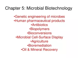
Chapter 15: Microbial Pathogenesis
Chapter 15: Microbial Pathogenesis. Microbial Pathogenesis Entry into the Host Must access and adhere to host tissues, penetrate or evade host defenses, and damage tissue to cause disease. Portals of Entry The three main portals of entry are: Mucous membranes Skin Parenteral.

Chapter 15: Microbial Pathogenesis
E N D
Presentation Transcript
Chapter 15: Microbial Pathogenesis
Microbial Pathogenesis Entry into the Host Must access and adhere to host tissues, penetrate or evade host defenses, and damage tissue to cause disease. Portals of Entry The three main portals of entry are: • Mucous membranes • Skin • Parenteral
Microbial Mechanisms of Pathogenicity: How Microorganisms Cause Disease
I. Mucous Membranes Epithelial tissue lining the: • Respiratory tract: Easiest and most frequently used entry site for microbes. • Gastrointestinal tract: Another common entry site. Enter through water, food, contaminated fingers and fomites. Must survive stomach HCl, enzymes, and bile. • Genitourinary tract: Entry site for most sexually transmitted diseases (STDs). • Conjunctiva: Membrane covering eyes and eyelids.
II. Skin Unbroken skin is impenetrable by most microbes. • Some microbes gain access through hair follicles and sweat glands. • Nectator americanus (hookworm) can bore through intact skin. • Certain fungi (dermatophytes) grow on skin and produce enzymes that break down keratin.
III. Parenteral Route Microbes are deposited directly into the tissues beneath the skin or mucous membranes. • Examples: Injections, bites, cuts, wounds, surgery, punctures, and splitting due to swelling or drying. Preferred Portal of Entry Many microbes have a preferred portal of entry which is a prerequisite to cause disease. • Example: Streptococcus pneumoniae that are inhaled can cause pneumonia; if swallowed generally don’t cause disease.
Number of Invading Microbes • Higher number of pathogens increase the likelihood of developing disease. • LD50: Lethal dose for 50% of hosts. Number of microbes that will kill 50% of inoculated test animals. • ID50: Infectious dose for 50% of hosts. Number of microbes that will cause a demonstrable infection in 50% of inoculated test animals.
Adherence • Attachment between of microbe to host tissue requires: • Adhesins or Ligands: Surface molecules on pathogen that bind specifically to host cell surface molecules. May be located on glycocalyx, fimbriae, viral capsid, or other surface structure. • Receptors: Surface molecules on host tissues to which pathogen adhesins bind. • Cell Wall Components • M protein: Found on cell surface and fimbriae of Streptococcus pyogenes. Mediates attachment an dhelps resist phagocytosis. • Waxes: In cell wall of Mycobacterium tuberculosis helps resist digestion after phagocytosis. • Enzymes • Extracellular enzymes (exoenzymes) lyse cells, form or dissolve clots, and dissolve materials in tissue. • . Leukocidins: Destroy white blood cells that are phagocytes. Produced by staphylococci and streptococci. • . Hemolysins: Destroy red blood cells. Produced by clostridium perfringens (gangrene) and streptococci. • . Coagulases: Produce blots in blood. Clots may protect bacteria from host immune system, by walling off site of infection. Produced by some staphylococci. • . Bacterial Kinases: Break down clots produced by body to isolate infection. Made by streptococci and staphylococci. • . Hyaluronidase: Breaks down hyaluronic acid which holds cells together in connective tissue. Made by some streptococci and gangrene causing clostridia. • . Collagenase: Breaks down collagen which forms connective tissue of muscles, skin, and other organs. Produced by several clostridia. • . Necrotizing Factors: Kill body cells. • . Hypothermic factors: Decrease body temperature. • . Lecithinase: Destroys plasma membrane of cells. • . Proteases: Break down proteins in tissue. • Penetration into Host Cells • Invasins: Surface proteins that alter actin filaments of host cell cytoskeleton, allowing microbes to enter cells. • Examples:Salmonella typhinurium and E. coli. • Cadherin: A glycoprotein that bridges junctions between cells, allowing microbes to move from one cell to another.
Microbial Mechanisms of Pathogenicity: How Microorganisms Cause Disease
How Bacterial Pathogens Penetrate Host Defenses Capsules • Increase the virulence of many pathogens. • Examples: Streptococcus pneumoniae, Klebsiella pneumoniae, Hemophilus influenzae, Bacillus anthracis, and Yersinia pestis. • Resist host defenses by impairing phagocytosis. • Host can produce antibodies to capsule, which attach to microbe and allow phagocytosis.
Cell Wall Components • M protein: Found on cell surface and fimbriae of Streptococcus pyogenes. Mediates attachment and helps resist phagocytosis. • Waxes: Cell wall of Mycobacterium tuberculosis helps resist digestion after phagocytosis.
Microbial Enzymes Extracellular enzymes (exoenzymes) lyse cells, form or dissolve clots, and dissolve materials in tissue. • Leukocidins: Destroy white blood cells that are phagocytes. Produced by staphylococci and streptococci. • Hemolysins: Destroy red blood cells. Produced by clostridium perfringens (gangrene) and streptococci. • Coagulases: Produce clots in blood, which may wall off site of infection from immune response. Produced by some staphylococci. • Bacterial Kinases: Break down clots produced by body to isolate infection. Made by streptococci and staphylococci. • Hyaluronidase: Breaks down hyaluronic acid which holds cells together in connective tissue. Made by some streptococci and gangrene causing clostridia.
Tissue Damage Caused by Microbial Enzymes of Clostridium perfringens Severe gangrene caused by Clostridium perfringens. Source: Tropical Medicine and Parasitology, 1997
Microbial Enzymes (Continued) • Collagenase: Breaks down collagen which forms connective tissue of muscles, skin, and other organs. Produced by several clostridia. • Necrotizing Factors: Kill body cells. • Hypothermic factors: Decrease body temperature. • Lecithinase: Destroys plasma membrane of cells. • Proteases: Break down proteins in tissue.
Tissue Damage Caused by Enzymes of Flesh-Eating Streptococcus pyogenes Necrotizing fasciitis with blood filled vesicles. Source: Perspectives in Microbiology, 1995
Penetration into Host Cells • Invasins: Surface proteins that alter actin filaments of host cell cytoskeleton, allowing microbes to enter cells. • Examples:Salmonella typhinurium and E. coli. • Cadherin: A glycoprotein that bridges junctions between cells, allowing microbes to move from one cell to another.
How Bacterial Cells Damage Host Cells Three mechanisms: • Direct Damage • Toxins* • Hypersensitivity Reactions * Most bacterial damage is carried out by toxins. 1. Direct Damage • Some bacteria can induce cells to engulf them (E. coli, Shigella, Salmonella, and Neisseria gonorrhoeae). • Microbial metabolism and multiplication kills host cells. • Other microbes enter the cell by excreting enzymes or through their own motility.
2. Toxin Production • Toxins: Poisonous substances produced by microbes. • Frequently toxins are the main pathogenic factor. • Toxigenicity: Ability of a microbe to produce toxins. • Toxemia: Presence of toxins in the blood. • Toxin effects: May include fever, cardiovascular problems, diarrhea, shock, destruction of red blood cells and blood vessels, and nervous system disruptions. • Of 220 known bacterial toxins, 40% damage eucaryotic cell membranes. • Two types of toxins: • Exotoxins • Endotoxins
A. Exotoxins • Proteins: Enzymes that carry out specific reactions. • Soluble in body fluids, rapidly transported throughout body in blood or lymph. • Produced mainly by gram-positive bacteria. • Most genes for toxins are carried on plasmids or phages. • Produced inside bacteria and released into host tissue. • Responsible for disease symptoms and/or death. • Cytotoxins: Kill or damage host cells. • Neurotoxins: Interfere with nerve impulses. • Enterotoxins: Affect lining of gastrointestinal tract. • Antibodies called antitoxins provide immunity. • Toxoids: Toxins that have been altered by heat or chemicals. Used as vaccines for diphtheria and tetanus.
Important Exotoxins • Diphtheria Toxin: Corynebacterium diphtheriae when infected by a phage carrying tox gene. Cytotoxin inhibits protein synthesis in eucaryotic cells. Two polypeptides: A (active) and B (binding). • Erythrogenic Toxins: Streptococcus pyogenes produces three cytotoxins which damage blood capillaries, causing a red rash. • Botulinum Toxins: Produced by Clostridium botulinum.Neurotoxin that inhibits release of neurotransmitter acetylcholine and prevents transmission of nerve impulses to muscles, causing flaccid paralysis. Extremely potent toxins. • Tetanus Toxin: Produced by Clostridium tetani. A neurotoxin that blocks relaxation of skeletal muscles, causing uncontrollable muscle spasms (lockjaw) and convulsions. • Vibrio Enterotoxin: Produced by Vibrio cholerae. Two polypeptides: A (active) and B (binding). The A subunit of enterotoxin causes epithelial cells to discharge large amounts of fluids and electrolytes. • Staphylococcal Enterotoxin: Staphylococcus aureus produces an enterotoxin similar to cholera toxin. Other enterotoxins cause toxic shock syndrome.
Rash of Scarlet Fever Caused by Erythrogenic Toxins of Streptococcus pyogenes
Muscle Spasms of Tetanus are Caused by Neurotoxin of Clostridium tetani Neonatal Tetanus (Wrinkled brow and risus sardonicus) Source: Color Guide to Infectious Diseases, 1992
Vibrio Enterotoxin Causes Profuse Watery Diarrhea Rice-water stool of cholera.The A subunit of enterotoxin causes epithelial cells to discharge large amounts of fluids and electrolytes. Source: Tropical Medicine and Parasitology, 1995
Diseases Caused by Staphylococcal Toxins Scalded Skin Syndrome Toxic Shock Syndrome
Endotoxins • Part of outer membrane surrounding gram-negative bacteria. • Endotoxin is lipid portion of lipopolysaccharides (LPS), called lipid A. • Effect exerted when gram-negative cells die and cell walls undergo lysis, liberating endotoxin. • All produce the same signs and symptoms: • Chills, fever, weakness, general aches, blood clotting and tissue death, shock, and even death. Can also induce miscarriage. • Fever: Pyrogenic response is caused by endotoxins.
Endotoxins (Continued) • Endotoxins do not promote the formation of effective antibodies. • Organisms that produce endotoxins include: • Salmonella typhi • Proteus spp. • Pseudomonas spp. • Neisseria spp. • Medical equipment that has been sterilized may still contain endotoxins. • Limulus amoebocyte assay (LAL) is a test used to detect tiny amounts of endotoxin.
Events leading to fever: • Gram-negative bacteria are digested by phagocytes. • LPS is released by digestion in vacuoles, causing macrophages to release interleukin-1 (IL-1). • IL-1 is carried via blood to hypothalamus, which controls body temperature. • IL-1 induces hypothalamus to release prostaglandins, which reset the body’s thermostat to higher temperature.
Shock: Any life-threatening loss of blood pressure. Septic shock: Shock caused by endotoxins of gram-negative bacteria. • Phagocytosis of bacteria leads to secretion of tumor necrosis factor (TNF), which alters the permeability of blood capillaries and causes them to lose large amounts of fluids. • Low blood pressure affects kidneys, lungs, and gastrointestinal tract.
Plasmids, Lysogeny, and Pathogenicity Plasmids: Small, circular pieces of DNA that are not connected to chromosome and are capable of independent replication. • R (resistance) factors contain antibiotic resistance genes. • Other plasmids contain genes for toxins and pathogenic factors: tetanus toxin, staphylococcal enterotoxin, E. coli enterotoxin (heat-labile), adhesins, and coagulase.
Bacteriophages: Can incorporate genetic material into chromosomal DNA and remain latent (lysogeny). Bacterial cell can change characteristics (lysogenic conversion) and produce certain toxins or pathogenic factors: • Diphtheria toxin • Capsules in S. pneumoniae • Botulinum neurotoxin • Staphylococcal enterotoxin • Cholera toxin.
Cytopathic Effects (CPE) of Viruses Viral infection may result in one or several of the following cytocidal or noncytocidal effects in infected cells: 1.Inhibit macromolecular synthesis (DNA, RNA, protein). Some viruses irreversibly stop mitosis (herpes simplex virus). 2.Release oflysosomalenzymes, resulting in cell death. 3. Inclusion bodies: Granules in cytoplasm or nuclei of infected cells. May contain viral parts. 4. Syncytium: Fusion of several adjacent cells to form a single giant cell.
Cytopathic Effects of Viruses (Cont.) 5. Metabolic changes in host without visibly damaging infected cells. May increase hormone or protein production by infected cells, which in turn affects other cells. 6. Interferon production: Interferon produced by infected cells, protects neighboring cells from infection. 7.Antigenic changes on cell surface, causing destruction of infected cells by immune system. 8. Chromosomal changes: Breakage and incorporation of oncogenes. 9. Transformation: Abnormal cells that have lost contact inhibition.
Microbial Mechanisms of Pathogenicity: How Microorganisms Cause Disease