Download
1 / 25
260 likes | 352 Vues
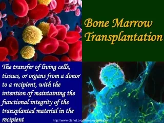
Explore the intricate mechanisms of hemopoiesis, stem cell functions, regulation, RBC structure, and system analysis in physiological and pathophysiological conditions. Includes methods of studying progenitors and hemopoietic systems. Dive into the roles of cytokines, lymphokines, growth factors, and tumor necrosis factors. Discover the unique structure and physiology of red blood cells, emphasizing hemoglobin protection, morphology, and life span.

E N D
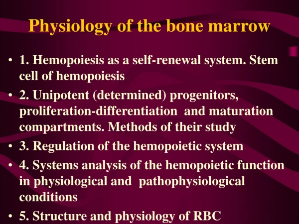
Physiology of the bone marrow • 1. Hemopoiesis as a self-renewal system. Stem cell of hemopoiesis • 2. Unipotent (determined) progenitors, proliferation-differentiation and maturation compartments. Methods of their study • 3. Regulation of the hemopoietic system • 4. Systems analysis of the hemopoietic function in physiological and pathophysiological conditions • 5. Structure and physiology of RBC
1. Hemopoiesis as a self-renewal system. • Stem cell of hemopoiesis • A self-renewal system loses mature (functional) cells steadily and replaces them continually. Normally it is composed of three cellular compartements: • - pluripotent stem cells which are capable of • - non declining (steady) autoreproduction • - differentiating into various developmental • lines (pluripotentiality) • - unipotent cells capable of dividing proliferation) • - differentiated functional (postmitotic) cells • unable of division • Blood forming organs represent a typical example of a self-renewal system (Fig. 1).
All non-stem populations are transitory. The transit times in the individual compartements: Two types of damage: - loss of mature cells (e.g., bleeding, inflammation) - damage to the stem cell and precursor compartement (e.g., radiation, cytotoxic drugs) Lymphatic organs are totally dependent on the marrow
Fig 2 : Erythropoiesis, the development and maturation of the red blood cells 2
2. Unipotent (determined) progenitors, proliferation-differentiation and maturation compartments. Methods of their study • The historically first method of the stem cell detection: colony forming units spleen (CFU-S) • Fig. 3, Fig. 4: The stages which cannot be differentiated • morphologically are detected by means of short- • term colonies in vitro
Besides, the stem and precursor cells could be identified by means of flow cytometry ( numbers of different cells) and light-activated cell sorting( preparing of homogenous cell populations, Fig. 5)
1. Regulation of the hemopoietic system Fig. 6: Hemopoietic inductive microenvironment(HIM) - fibroblast-type “reticulum” cell – granulopoietic islands - macrophage-type “reticulum” cell – erythroblastic islands 6
Cytokines = peptides taking part in the signalling among the immune and hematopoietic system cells. No hormones (endocrine). Types: • - Interleukins (Il-1, 2,...etc.) • MF, T cells, stromal cells (HIM) growth • and differentiation of • - lymphocytes • - hematopoetic stem cells (some of them • are known as “colony stimulating • factors”, CSF) • Ils taking part in steady-state hemopoiesis: • - multi-CSF (= Il-3) from helper T-cells • - GM-CSF • - Il-6 • - Il-7 (development of B- and T-lymphocytes)
In non-steady-state-conditions: production • of a number of growth factors more specific in • their actions which are usually produced by • activated blood cells or fibroblasts: Il-4 and Il-9 • (production of basophils and mast cells), Il-5 • (eosinophils), G-CSF, M-CSF, EPO • - Interferons: induced in response to a variety of • agents including viruses, microorganisms and • endotoxins. Upon induction, they circulate to • neighboring cells which they stimulate to make • antiviral proteins
Lymphokines: Proteins secreted by some helper T cells after they are primed by contact with an antigen. Are not antibodies but are mediators of cellular immunity. They activate various white blood cells, incl. other lymphocytes. Examples: interleukin 2, some interferons, migration inhibition factor (MIF) • Tumor necrosis factors: have cytotoxic effects on tumor cells but not on normal cells. TNF is secreted by macrophages in response to bacterial infection and other challenges, TNF by helper T lymphocytes and cytotoxic lymphocytes. Synergistic with interferons
4. Systems analysis of the hemopoietic function in physiological and pathophysiological conditions • The homeostasing of the blood cell production is regulated by several feedbacks. Their interplay may be rather complex and can be understood by means of mathematical modelling only (Fig. 7, 8, 9)
5. Structure and physiology of RBC • The RBS have lost • - mitochondria, citric acid cycle and oxydative • phosphorylation forming ATP anaerobic • glycolysis must be the main source of ATP in • them; • - ribosomes no protein synthesis, detrition of • enzymes etc. • RBC membrane: • Fig. 10: glycophorins can attach lectins = phytohemagglutinins, viruses and malaria parasites
Fig. 11a, b: Spectrin and actin filamentous network responsible for the biconcave shape of RBC; ankyrinlinks spectrin molecules to anion-transportproteins (“band 3”)