Download
1 / 36
571 likes | 2.86k Vues
BIOLOGICAL EFFECTS OF IONIZING RADIATION AT MOLECULAR AND CELLULAR LEVELS. Module VIII-a. Historical b ackground. Discovery of X r ays (1895). Wilhelm Conrad Roentgen. Discovery of u ranium ’s n atural r adioactivity. Antoine Henri Becquerel. Marie Curie.

E N D
BIOLOGICAL EFFECTS OF IONIZING RADIATION AT MOLECULAR AND CELLULAR LEVELS Module VIII-a
Historical background Module Medical VIII.
Discovery of X rays (1895) Wilhelm Conrad Roentgen Module Medical VIII.
Discovery of uranium’snatural radioactivity Antoine Henri Becquerel Marie Curie Module Medical VIII.
First reports on harmful effects of radiation • First radiation-induced skin cancer reported • in 1902 • First radiation-induced leukemia described • in 1911 • 1920s:bone cancer among radium dial painters • 1930s:liver cancer and leukemia due to Throtrast administration • 1940s: excess leukemiaamong first radiologists Module Medical VIII.
Studies of Japanese A-bomb survivors Module Medical VIII.
Effects of radiation on cells at atomic level Ionization Excitation Module Medical VIII.
Mechananisms of damage at molecular level Module Medical VIII.
Direct action Ionizing radiation + RH R- + H+ Bond breaks OH I R – C = NH imidol (enol) O II R – C = NH2 amide (ketol) Tautomeric Shifts Module Medical VIII.
Indirect action OH- H O H+ Xray ray e- H Ho P+ OHo Module Medical VIII.
Radiolysis of H2O molecule Shared electron Shared electron H-O-H H+ + OH- (ionization) H-O-H H0+OH0 (free radicals) Module Medical VIII.
Reactions with free radicals H0 + OH0 HOH (recombination) H0 + H0 H2 (dimer) OH0 +OH0 H2O2 (hydrogen peroxide) OH0+RH R0+HOH (radical transfer) Module Medical VIII.
Effects of oxygen on free radical formation Oxygencan modify the reaction by enabling creation of other free radical species with greater stability and longer lifetimes H0+O2 HO20 (hydroperoxy free radical) R0+O2 RO20 (organic peroxy free radical) Module Medical VIII.
Lifetimes of free radicals RO2o HO2o Ho OHo 3nm OHo Ho Because short life of simple free radicals (10-10sec), only those formed in water column of 2-3 nm around DNA are able to participate in indirect effect Module Medical VIII.
Relation between LET and action type Direct action is predominant with high LET radiation, e.g. alpha particles and neutrons Indirect action is predominant with low LET radiation,e.g. X and gamma rays Module Medical VIII.
Biochemical reactions with ionizing radiation DNA is primary target for cell damage from ionizing radiation Module Medical VIII.
Types of radiation induced lesions in DNA Base damage Single-strand breaks Double strand breaks Module Medical VIII.
Mechanisms of DNA repair Module Medical VIII.
DNA restoration failure Incorrect repair of DNA damage Unrejoined DNA double strand breaks Cytotoxic effect Mutations Module Medical VIII.
Chromosomes Module Medical VIII.
DNA lesions and chromosome aberrations DNA SİNGLE STRAND BREAK DNA DOUBLE STRAND BREAK Module Medical VIII.
Radiation induced chromosomal aberrations Module Medical VIII.
Effect of radiation on cell Cell kinetics Module Medical VIII.
Radiosensitivity of cell in cell cycle Relative Survivability G1 S G2 G1 M Relative survivability of cells irradiated in different phases of the cell cycle. Synchronised cells in late G2 and in mitosis (M) showed greatest sensitivity to cell killing. Module Medical VIII.
Mitotic death NORMAL IRRADIATED Module Medical VIII.
Bergonié and Tribondeaus’ ‘law’ (1906) • The most ‘radiosensitive’ cells are • actively proliferating/dividing at the time of exposure • undifferentiated (non-specialized in structure and function) Module Medical VIII.
Interphase death Why are peripheral blood lymphocytes highly sensitive to radiation, although well differentiated? Module Medical VIII.
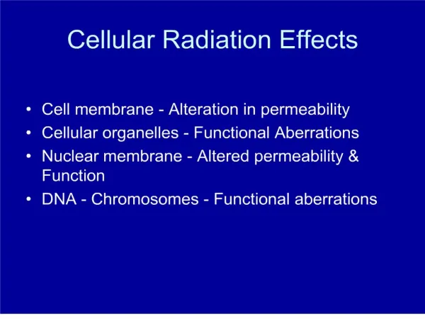
Radiation induced membrane damage Module Medical VIII.
Modification of radiation injury • Dose rate and fractionation • Radiation quality • Temperature • Chemical modification • Oxygen • Radiosensitizing agents • Radioprotective agents Module Medical VIII.
Dose rate and fractionation Time Acute dose Acute exposure with high dose rate Time Prolonged exposure with lower dose rate Fractionated dose Module Medical VIII.
Radiation quality Module Medical VIII.
Survival curve for mammalian cells exposed to high- (A) and low-LET (B)radiation n Dq 1-1/e 1-1/e ,037 D0 D0 B A Module Medical VIII.
Temperature • For cell kiling effects, tissues are more radiosensitive at higher temperatures • Chromosome aberrations increase at lower temperatures (suppression of repair process) Module Medical VIII.
Chemical modification: oxygen • Dissolved oxygen in tissues increases stability and toxicity of free radicals • Oxygen enhancement ratio (OER) is determined by: The OER has a maximum value of 3.0 Dose required to cause effect without oxygen OER = Dose required to cause effect with oxygen Module Medical VIII.
Radiosensitizing agents • Halogenated and substituted analoges of DNA bases:5-bromo-uracil and 6-thio-guanine • Electroaffinic compounds: Nitroimidazoles(misonidazole, nitroimidazole, and nitrofuran)sensitization enhancement ratio (SER) of 1.2 to 1.4 Module Medical VIII.
Radioprotective agents • Thiols(cysteine, 2-mercaptoethylamine, cystamine and thiourea). Thiols have dose reduction factor (DRF) ratio of 1.4 to 2.0 They are thought to protect cells by • scavenging free radicals • producing hypoxia • temporarily inhibiting DNA synthesis, allowing time for the repair enzymes to complete repair of sublethal damage • forming disulphide bonds in proteins,thereby strengthening them Module Medical VIII.