Download
1 / 9
130 likes | 893 Vues
Preoperative radiograph of the mandibular left premolar (35). Note the wide-open apex and periapical radiolucent lesion. Working length radiograph. Resorbable collagen sponge to create a periapical barrier fro the compaction of the Portland cement. White Portland cement + distilled water.

E N D
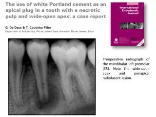
Preoperative radiograph of the mandibular left premolar (35). Note the wide-open apex and periapical radiolucent lesion.
Resorbable collagen sponge to create a periapical barrier fro the compaction of the Portland cement
Preoperative radiograph with white Portland cement placed at the apical portion of the canal (approximately 3 mm).
Seven months later The patient was instructed to return 1 week later for the continuation of treatment but he did not return as planned. Seven months after the initial intervention the patient returned to complete the treatment.
Verifying the integrity of the Portland cement plug with a size 20 file.
Immediate postoperative radiograph with root-canal filling and the white Portland cement in the apical third.