Download
1 / 43
680 likes | 2.01k Vues
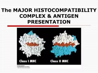
The MAJOR HISTOCOMPATIBILITY COMPLEX & ANTIGEN PRESENTATION. MHC. MHC - tightly linked cluster of genes whose products are associated with intracellular recognition and self/nonself discrimination Major role in determining whether transplanted tissue will be histocompatible or histoincompatible

E N D
MHC • MHC - tightly linked cluster of genes whose products are associated with intracellular recognition and self/nonself discrimination • Major role in determining whether transplanted tissue will be histocompatible or histoincompatible • Central role in development of humoral and cell-mediated immune response • Critical role in antigen recognition by T cells
General Organization and Inheritance of MHC • Gorer and Snell (1930’s) concept of rejection of foreign tissue is an immune response to cell surface molecules • Snell (1980)- Nobel Prize
Location and Function of MHC Regions • Collection of genes within a long stretch of DNA on chromosome 6 in humans and chromosome 17 in mice • MHC - Human Leukocyte Antigen (HLA) • H-2 Complex (mice) • MHC genes organized in regions encoding 3 classes of molecules
Class I MHC Genes • MHC Class 1 mediates immune responses against endogenous antigens, antigens that are already found in the cell. • Usually, these cells that are expressing MHC class 1 are viral-infected or are tumor cells. • MHC Class 1 presents peptides that are 8 – 10 amino acids in size, which will then be recognized by the cytotoxic T cells. • MHC Class 1 is found on all nucleated cells.
Class II MHC Genes • MHC class 2 mediates immune responses against exogenous antigens, antigens that are found outside of the cell, in the cytosol. • MHC class 2 will bind with amino acid residues that are 13 – 18 in size and will be recognized by T helper cells. • The MHC class 2 protein is found on cells like the B lymphocytes, macrophages, monocytes, dendritic cells, and endothelial cells. • These cells are phagocytic and can engulf an extracellular antigen.
Class III MHC Genes • Generally encode secreted proteins associated with the immune process • Reason for location within the MHC region is uncertain
Haplotype- set of genes located on a single chromosome and the characteristics dependent on them An individual has 2 haplotypes of each set of genes (maternal/paternal). MHC genes expressed codominantly (both maternal and paternal products expressed in same cells MHC Haplotypes
Class I Structure • Contains large chain associated with a smaller 2-microglobulin molecule • chain is a polymorphic transmembrane glycoprotein (45 kDa) • 2- microglobulin molecule is an invariant protein (12kDa) encoded by a gene on a different chromosome • Association of chain with 2 microglobulin is required for expression of Class I on cell membranes
Class I Structure • chain anchored in plasma membrane by its hydrophobic transmembrane segment & hydrophilic cytoplasmic tail • chain has 3 external domains • Homology between 3 & 2 microglobulin & constant regions in immunoglobulins • Peptide-binding cleft
Class II Structure • Contains 2 different polypeptide chains: chain (33 kDa) & a chain (28 kDa) • Each chain has 2 external domains • Antigen binding cleft for processed antigens • heterodimer “dimer of dimers”
Class I MHC-Peptide Interaction • Class I bind peptides & present these peptides to CD8+ T-cells • Each type of Class I (A,B,C in humans K,D,L in mice) bind unique set of peptides • Single nucleated cell expresses 105 copies of each Class I molecule • Many different peptides will be expressed simultaneously on the surface by Class I MHC • Endogenous processing pathway
Class II MHC-Peptide Interaction • Class II MHC binds peptides and presents these peptides to CD4+ T cells • Can bind of variety of peptides • Endocytic processing pathway
Polymorphism of Class I & II MHC • Polymorphism - presence of multiple alleles at a given genetic locus within a species • Diversity of MHC within a species results from polymorphism • MHC expresses by an individual does not change over time but they may differ significantly from those expressed by another individual of the same species • 1012 theoretical diversity of mice in each Class I & II MHC gene
Polymorphism of Class I & II MHC • MHC also serve as antigens to let your immune system know what is self and what is non-self. • The different allele combinations make up the identity of your MHC. • Within each of these genes, there are many alleles. • The main point is that there are lots and lots of alleles and thousands of combinations, which is why finding someone to whose HLA markers match up w/ another person is so difficult. • But it’s not impossible, there are thousands of transplants every year.
Class III MHC Molecules • Several structurally & functionally diverse proteins encoded within the 3rd region of MHC • Includes several complement components, tumor necrosis factors ( & ), 2 heat shock proteins • Not membrane proteins and have no role in antigen presentation, although most play a role in immune response
Heat Shock Proteins • Unusual group of highly conserved proteins that are produced by cells in response to various stresses including heat shock, nutrient deprivation, oxygen radicals, and viral infection • Linked to certain autoimmune diseases
Antigen Processing & Presentation • Formation of peptide-MHC complexes require that a protein antigen be degraded into peptides & displayed within the cleft of the MHC molecule on the cell membrane. The sequence of the above events is called antigen processing. • The display of the transported peptide-MHC molecules on the cell membrane is called antigen presentation.
Antigen Processing & Presentation (cont.) • Class I MHC molecules bind peptides derived from endogenous antigens processed in the cytoplasm. • Class II MHC molecules bind peptides derived from exogenous antigens that are internalized by phagocytosis or endocytosis & processed within the endocytic pathway.
Self-MHC Restriction of T cells • Zinkernagel & Doherty demonstrated the self-MHC restriction of CD8+ T cells by immunizing mice with Lymphocyte choriomeningitis (LCM) virus. • TC cells only killed syngeneic virus-infected target cells, showing that TC cell & the virus-infected target cell must share class I molecules encoded by K or D MHC regions.
Classic experiment of Zinkernagel and Doherty • They demonstrate that antigen recognition by TC cells exhibits MHC restriction. • H-2k mice were primed with the lymphocytic choriomeningitis (LCM) virus to induce cytotoxic T lymphocytes (CTLs) specific for the virus. • Spleen cells from this LCM-primed mouse were then added to target cells of different H-2 haplotypes that were intracellularly labeled with 51Cr (black dots) and either infected or not with the LCM virus. • CTL-mediated killing of the target cells, as measured by the release of 51Cr into the culture supernatant, occurred only if the target cells were infected with LCM and had the same MHC haplotype as the CTLs.
Self-MHC Restriction of T cells (cont.) • CD4+ & CD8+ cells can only recognize antigen when presented on the membrane of a cell by a self-MHC molecule (self-MHC restriction). • Rosenthal & Shevach showed that antigen –specific proliferation of TH cells only occurred in response to antigen presented by macrophage of the same MHC haplotype. • These results indicate that the CD4+ TH cell can proliferate in response to antigen presented by macrophage that shared MHC alleles. - CD4 T cells are class II MHC restricted.
Experimental demonstration of self-MHC restriction of TH cells. Peritoneal exudate cells from strain 2, strain 13, or (2 13) F1 guinea pigs were incubated in plastic Petri dishes, allowing enrichment of macrophages, which are adherent cells. The peritoneal macrophages were then incubated with antigen. These “antigen-pulsed” macrophages were incubated in vitro with T cells from strain 2, strain 13, or (2 13) F1 guinea pigs, and the degree of T-cell proliferation was assessed. The results indicated that TH cells could proliferate only in response to antigen presented by macrophages that shared MHC alleles.
Antigen Presenting Cells • Cells expressing class I or II MHC molecules can present peptides to T cells. • By convention, cell that display peptides associated with class I MHC molecules to CD8+ T cells are referred to as target cells. • Those cells that display peptides associated with MHC class II molecules to TH cells are called antigen presenting cells.
Antigen Processing • Extracellular (exogenous) antigens are eliminated by secreted antibody whereas intracellular (endogenous) antigens are eliminated by CTLs. • There are 2 different antigen-presenting pathway to mediate responses. - Endocytic pathway - Cytosolic pathway
Endocytic (exogenous)Pathway • APCs can internalize antigen by phagocytosis &/or endocytosis. • Macrophages do both; B cells use receptor-mediated endocytosis. • After antigen is internalized, it is degraded into peptides.
Endocytic Pathway (cont.) • Internalized antigen takes 1-3 hours to traverse the endocytic pathway & appear on cell membrane in the form of peptide-class II MHC complexes. • Internalized antigen moves from early to late endosomes & finally to lysosomes where they are hydrolyzed into oligopeptides of about 13-18 residues that bind to class II MHCs.
Exogenous Pathway • The pathway begins by phagocytosis by the cell of a foreign agent, an organism, bacteria, etc… • The antigen is now in a phagosome. A lysosome will fuse with the phagosome to become a phagolysosome. • The antigen will be degraded into smaller peptides. • With the help of sorting signals from the invariant chain (that’s attached to the MHC class 2), the MHC class two will migrate to the phagolysosome, where it will bind to components that are 13 – 18 amino acids in size. Once bound, the MHC class 2 will migrate to the membrane to display the antigen. • A helper T cell will recognize the complex and trigger the appropriate response, such as secreting cytokines and chemokines to control whatever kind of infection is taking place.
Endogenous Pathway • We start with an antigen that’s already in the cell. It will be broken down into smaller peptides by a protease. • The peptides will be transported into the endoplasmic reticulum where MHC class 1 is located. • The 8 – 10 amino acid residues will bind with MHC class 1 and once that happens, the MHC class 1 and antigen will migrate to the cell surface, where it will present the antigen. • Cytotoxic T cells will recognize this complex and initiate the appropriate immune response to kill this cell.
Cytosolic Pathway • Endogenous antigens are degraded into peptides that can be presented in class I MHC molecules to TC cells involving similar mechanisms as of intracellular proteins. • Ubiquitin → Ubiquitin-protein conj → Proteosome • Subunits of large cytoplasmic proteolytic complex are called low-molecular mass polypeptides (LMP).
Necrotizing granulomatous lesions in TAP-deficiency syndrome