Download
1 / 79
1.14k likes | 3.18k Vues
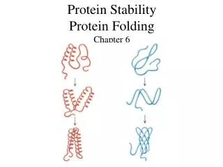
Protein Stability Protein Folding Chapter 6. Protein Stability. Protein stability is the net balance of forces, which determine whether a protein will be in its native folded conformation or a denatured state.

E N D
Protein Stability • Protein stability is the net balance of forces, which determine whether a protein will be in its native folded conformation or a denatured state. • Protein stability normally refers to the physical (thermodynamic) stability, not the chemical stability.
Chemical Stability • Chemical stability involves loss of integrity due to bond cleavage. • deamination of asparagine and/or glutamine residues, • hydrolysis of the peptide bond of Asp residues at low pH, • oxidation of Met at high temperature, • elimination of disulfide bonds • disulfide interchange at neutral pH • Other processes include thiol-catalyzed disulfide interchange and oxidation of cysteine residues.
Protein Stability • The net stability of a protein is defined as the difference in free energy between the native and denatured state: • Both GN and GU contribute to G • The free energy may be readily calculated from the following relationships: K = [N]/[U] = FN/(1- FN), FN = fraction folded DG = GN - GU = -RTlnK • Decreasing the energy of the folded state or increasing the energy of the unfolded state have the same effect on DG.
Protein Stability • Protein stability is important for many reasons: • Providing an understanding of the basic thermodynamics of the process of folding, • increased protein stability may be a multi-billion dollar value the in food and drug processing, and in biotechnology and protein drugs. • Two relatively recent innovations, which have had major impact in the study of the thermodynamics of proteins were the development of very sensitive techniques, differential scanning calorimetry (especially by Privalov and Brandts) and site-directed mutagenesis.
Stability of the Folded State • Measuring protein stability is measuring the energy difference between the U (unfolded) and F (folded) states. • The average stability of a monomeric small protein is about 5 - 10 kcal/mol, which is very small! DG = GN - GU = -RTlnK K=e-DG/RT = e-10x1000/(2x298) =2x 10 7 • i.e. in aqueous solution, at room temperature, the ratio of folded : unfolded protein is 2x 10 7 : 1!
Stability of the Folded State • K as the equilibrium constant, is the ratio of the forward (f) and the reverse (u) rate constant. K=kf/ku • If a typical protein refolds spontaneously with a rate constant of kf = 1 s-1, its rate of spontaneously unfolding under the same condition will be 10-7 s-1.The half life is 0.693/10-7 s = 80 days. • This suggests that the unfolding of proteins will only be transient. • We have to perturb the equilibrium to enable us to measure the unfolding of proteins using urea, pH, etc.
Techniques for Measuring Stability • Any methods that can distinguish between U and F Absorbance (e.g. Trp, Tyr) Fluorescence (Trp)-difference in emission max & intensity. CD (far or near UV) - (2o or 3o) NMR DSC (calorimetry) Urea gradient gels - difference in the migrating rates between F and U. Catalytic activity Chromophoric or fluorophoric probes
Denaturing Proteins at Extreme pHs • High pH and low pH denature many, but not all proteins (many are quite stable at pH 1!). • The basic idea is that the net charge on the protein due to the titration of all the ionizing groups leads to intramolecular charge-charge repulsion, which is sufficient to overcome the attractive forces (mostly hydrophobic and dispersive) resulting in at least partial unfolding of the protein. • The presence of specific counterion binding leads to formation of compact intermediate states such as the molten globule (substantial secondary structure, little or no tertiary structure, relatively compact size compared to the native state).
Denaturants • The effects of denaturants such as urea (usually 8 M) or Guanidinium Hydrochloride (usually 6 M GuHCl) are complex, and currently are best thought of as involving preferential solvation of the denatured (unfolded) state, involving predominantly hydrophobic related properties, and to a lesser extent H-bonding (both side-chains and backbone appear to be more soluble in the presence of the denaturants). • There is no a very good solvent because solvents that are good for the hydrophobic components are bad for the hydrophilic ones and vice versa. • As in the case of pH-induced denaturation, not all proteins are unfolded by these denaturants. • Protein stability: SCN- < Cl- < Urea < SO4 2- e. g. midpoints of unfolding transition for RNase: GuSCN = 0.3M, GuHCl = 0.8 M, and urea nearly 3 M.
Two-state Unfolding of Protein • Keq=[N]/[U]= ( [θ]obs- [θ]D)/( [θ]N- [θ]D) = FN/(1- FN) FN = fraction folded
Denaturants • It is common to extrapolate the data for the unfolding transition as a function of denaturant to 0 M to give the value in water (e.g. G(H2O)). DG D-N = DG H20D-N - m D-N [denaturant] DG H20D-N is about –5 to –10 kcal/mol • The extrapolation can have large errors.
m - value • m-value reflects the dependence of the free energy on denaturant concentration • Typically for urea m ~ 1 kcal/mol • For GuHCl m ~ 3 kcal/mol • The variation in slope (m) is believed to be due to change in the solvent accessible area of hydrophobic residues. The m-value is related to how cooperative the transition is, how much structure remains in the denatured state, perhaps how much denaturant binds to the unfolded state, etc. • It’s important to note that because of different values of m, two proteins that have Cm is such that one may appear more stable, but, in fact, the opposite is true in the stability (based on DG H20D-N).
Thermal Denaturation • The effects of temperature on protein structure have been, and are, controversial, since most proteins can show the phenomenon of cold denaturation, under appropriate conditions! • Disruption of hydrogen bonding and increasing hydrophobicity occurs with thermal denaturation.
Differential Scanning Calorimetry (DSC) • DSC measures the heat required to raise the temperature of the solution of macromolecules relative to that required to the buffer alone (heat obtained by substracting two large numbers). • DSC can be used to directly measure the enthalpy and melting temperature of a thermally induced transition. At Tm (50% unfolded), DG = 0, DH = TDS
Thermal Denaturation • It is generally assumed that Cp is constant with respect to temperature. However, Privalov observed that that Cp was positive for denaturation, i.e. the heat capacity Cp was greater for the unfolded state than the folded state. Cp = H/T = TS/T • It is probably the change in ordered water structure between the native and denatured states which accounts, at least in part, for the change in Cp.
Thermal Denaturation • The Van't Hoff eq: dlnK/d(1/T) = -H/R • Van't Hoff plots (lnK vs. 1/T) of the thermal denaturation of proteins are non-linear, indicating that H varies with temperature. • This implies that the heat capacity for the folded and unfolded proteins are different! DH/DT = Cp = (CpU - CpN) • Since H = Ho + Cp(T-To), S = So + Cp ln(T/To) and G(T) = Ho - T So + Cp [(T - To) - Tln(T/To )] where T0 is any reference temperature (usually set = Tm). • The Gibbs Helmholz equation. G(T) = Hm(1-T/Tm) - Cp[(Tm - T) + Tln(T/Tm)] • The temperature where S = 0, Ts = Tm exp(-Hm/[TmCp])
Thermal Denaturation • There are two important forms of enthalpy as far as protein unfolding is concerned, • the Van't Hoff enthalpy, from the temperature dependence of the equilibrium constant, DHVH, • and the enthalpy measured calorimetrically (the area under the peak), DHcal. • If these are equal, it means there are no populated intermediates present at the Tm, i. e. the system is a two-state one. • For most proteins DHVH/Dhcal = 1.05 ± 0.03 for two-state.
Thermophilic Proteins • Living organisms can be found in the most unexpected places, including deep sea vents at > 100 ºC and several hundred bars pressure, in hot springs, and most recently, deep in the bowels of the earth, living off H2 formed by chemical decomposition of rocks! • The proteins found in thermophilic species are much more stable than their mesophilic counterparts (although this corresponds to only 3 - 8 kcal/mol of free energy). • However, the overall three-dimensional structures will be essentially the same for both thermophilic and mesophilic proteins. • It only takes stability of a couple of H-bonds, you can understand why there are no gross differences in structure between thermophilic and mesophilic proteins. • The upper limit of temperature growth for bacteria is about 110 º C. • Many of the species found in these extreme environments (T > 100C, pH 2) belong to the Archeae kingdom.
Thermophilic vs Mesophilic Proteins • Thermophilic proteins have increased amounts of Arg, increased occurrence of Ala in helices, and Gly/Ala substitutions (which affect the entropy of the denatured state, and thus its free energy) and increased number of salt bridges. • Each of these alone makes only a small effect, but several such changes are enough. In general, it appears that there is no single determinant of increased thermal stability; each protein is a unique case, typically involving variations in hydrophobic interactions, H-bonds, electrostatic interactions, metal-ligand (e. g. Ca2+) binding, and disulfide bonds. There is some suggestion that better packing may also play a role.
Stability-activity Trade-off? • Some enzymes from thermophiles that are very stable at normal temperatures have low activities at the lower temperatures. • There are is a compromise between the stability and activity in the structure of the active site of a protein. • There are several positions in the active site can be mutated to give more stable but less active protein. • Activity can then be increased further at an unacceptable expense to stability. • Active site of enzymes and binding sites of proteins are a general source of instability, because they contain groups that are exposed to solvent in order to bind substrates and ligands, and so are not paired with their normal types of partners.
Aldehyde Ferredoxin Oxidoreductase • The crystal structure of an unusual hyperthermophilic enzyme, aldehyde ferredoxin oxidoreductase, a tungsten-containing enzyme, has been solved. • The optimum temperature for this enzyme is > 95 C!! The amino acid composition is close to the average for all prokaryotic proteins except glutamine. It is 45 % helical, 14 % sheet. There are no disulfide bonds. • As observed with many other thermophilic proteins there may be an increased number of salt bridges. • What may be significant is that the solvent accessible area is reduced, although the fraction of polar/hydrophobic is similar to other proteins.
Cold Denaturation • The free energy curve starts to drop at lower temperatures as predicted by the thermodynamics of protein folding. • In the past few years, several proteins have been shown to exhibit cold denaturation under destabilizing conditions, in usually either low pH or moderate denaturant concentration. • Fink, A. L. observed a cold Denaturation for a Staphylococcal Nuclease Mutant under neutral pH and no-denaturant conditions.
Factors Affecting Protein Stability • 1) pH: proteins are most stable in the vicinity of their isoelectric point, pI. In general, electrostatic interactions are believed to contribute to a small amount of the stability of the native state; however, there may be exceptions. • 2) Ligand binding: It has been known for a long time that binding ligands, e.g. inhibitors to enzymes, increases the stability of the protein. This also applies to ion binding --- many proteins bind anions in their functional sites.
Factors Affecting Protein Stability • 3) Disulfide bonds: It was observed that many extracellular proteins contained disulfide bonds; whereas intracellular proteins usually did not exhibit disulfide bonds. • In addition, for many proteins, if their disulfides are broken (i.e. reduced) and then carboxymethylated with iodoacetate, the resulting protein is denatured, i.e. unfolded, or mostly unfolded. • Disulfide bonds are believed to increase the stability of the native state by decreasing the conformational entropy of the unfolded state due to the conformational constraints imposed by cross-linking (i. e. decreasing the free energy of the unfolded state). Most protein have "loops" introduced by disulfides of about 15 residues, but rarely more than 25.
Factors Affecting Protein Stability • 4) Not all residues make equal contributions to protein stability. In fact, it makes sense that interior ones, inaccessible to the solvent in the native state, should make a much greater contribution than those on the surface, which will also be solvent accessible in the unfolded state. • Proteins are very malleable, i.e. a mutation at a particular residue tends to be accommodated by changes in the position of adjacent residues, with little further propagation.
Denatured States • If the denatured state involves most residues in a fully extended peptide chain conformation, i. e. maximal solvent exposure, then substitutions involving solvent-exposed residues in the native state will have limited effect. • If, on the other hand, the denatured state have considerable residual structure, then it is also possible that mutations may affect the conformation and free energy of the unfolded state; in extreme cases, perhaps only the denatured state and not the native state!
wt m+ m- m-value • The m-value changes can be used to understand the nature of denatured state. • The effect of mutations to the protein stability can be estimated using the change of DG H20D-N • For some of the mutation, the m-value is changed. The different m-values related to the difference between the number of molecules of solvent bound in the native vs. denatured state. Since for the folded stated we have similar structure, the number of solvent molecules bound to the folded state is about the same, and the m-value difference reflects the different distribution of denatured state. • => more or less exposure of hydrophobic residues.
Different Unfolded States m+ mutant has a more exposed unfolded state than that of m- mutant. m+ mutant M- mutant smallest
Protein Folding • Protein folding considers the question of how the process of protein folding occurs, i. e. how the unfolded protein adopts the native state. • This has proved to be a very challenging problem. It has aptly been described as the second half of the genetic code, and as the three-dimensional code, as opposed to the one-dimensional code involved in nucleotide/amino acid sequence. • Predict 3D structure from primary sequence • Avoid misfolding related to human diseases • Design proteins with novel functions
Anfinsen Experiment • Denaturation of ribunuclease A ( 4 disulfide bonds) with 8 M Urea containing b-mercaptoethanol to random coil, no activity
Anfinsen Experiment • After renaturation, the refolded protein has native activity despite the fact that there are 105 ways to renature the protein. • Conclusion: All the information necessary for folding the peptide chain into its native structure is contained in the primary amino acid sequence of the peptide.
Anfinsen Experiment • Remove b-mercaptoethanol only, oxidation of the sulfhydryl group, then remove urea → scrambled protein, no activity • Further addition of trace amounts of b-mercaptoethanol converts the scrambled form into native form. • Conclusion: The native form of a protein has the thermodynamically most stable structure.
The Levinthal Paradox • There are vastly too many different possible conformations for a protein to fold by a random search. • Consider just for the peptide backbone, there are 3 conformations per amino acid in the unfolded state, For a 100 a.a. protein we have 3100 conformations. • If the chain can sample 1012 conformations/sec, it takes 5 x 1035 sec (2 x 1028 year) • Conclusion: Protein folding is not random, must have pathways.
Equilibrium Unfolding • switch off part of the interactions in the native protein under different denaturing conditions such as chemical denaturants, low pH, high salt and high temperature • understand which types of native structure can be preserved by the remaining interactions
Equilibrium Unfolding • Using many probes to investigate the number of transitions during unfolding and folding • For 2-state unfolding, all probes give the same transition curves. Single domains or small proteins usually have two-state folding behavior. • For 3-state unfolding, there are more than one transitions or different probes have different transition curves
Molten Globule State (MG) • It is an intermediate of the folding transition U→MG→F • It is a compact globule, yet expanded over a native radius • Native-like secondary structure, can be measured by CD and NMR proton exchange rate • It has a slowly fluctuating tertiary structure which gives no detectable near UV CD signal and gives quenched fluorescence signal with broadened NMR chemical peaks • Non-specific assembly of secondary structure and hydrophobic interactions, which allows ANS to bind and gives an enhanced ANS fluorescence • MG is about a 10 % increase in size than the native state
Fluorescence A. 1 - native 3 - MG 2,4 - unfolded B. 1 - native 3,4 - MG 2 - unfolded
Kinetic Folding Pathways • U→ I →II → N • Not all steps have the same rate constants. • Intermediates accumulate to relatively low concentrations, and always present as a mixture • Identify kinetic intermediates • Measuring the rate constants • Figure out the pathways • Slow folding • Formation of disulfile bond • Pro isomerization
Unfolded State • The unfolded state is an ensemble of a large number of molecules with different conformations.
Three Classic Models of Protein Folding • The Framework model proposed that local elements of native local secondary structure could form independently of tertiary structure (Kim and Baldwin). These elements would diffuse until they collided, successfully adhering and coalescing to give the tertiary structure (diffusion-collision model)(Karplus & Weaver).
The classic Nucleation Model • The classic nucleation model postulated that some neighboring residues in the sequence would form native secondary structure that would act as a nucleus from which the native structure would propagate, in a stepwise manner. Thus, the tertiary structure would form as a necessary consequence of the secondary structure (Wetlaufer).