CHAPTER 6 Enzymes
CHAPTER 6 Enzymes. 6.1 An Introduction to Enzymes Catalysts, Proteins, Reaction Types 6.2 How Enzymes Work Rate Enhancement, Activation energy, Catalytic Mechanism 6.3 Enzyme Kinetics Steady-State, Michaelis-Menten Formalism Inhibition 6.4 Examples of Enzymatic Reactions

CHAPTER 6 Enzymes
E N D
Presentation Transcript
CHAPTER 6Enzymes 6.1 An Introduction to Enzymes • Catalysts, Proteins, Reaction Types 6.2 How Enzymes Work • Rate Enhancement, Activation energy, Catalytic Mechanism 6.3 Enzyme Kinetics • Steady-State, Michaelis-Menten Formalism • Inhibition 6.4 Examples of Enzymatic Reactions 6.5 Regulatory Enzymes
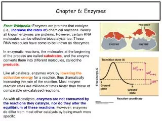
6.1 What are Enzymes? Enzymes are catalytically active biological macromolecules Specific, Efficient, Active in Aqueous Solution Most enzymes are globular proteins, however some RNA (ribozymes, and ribosomal RNA) also catalyze reactions Non-peptide Co-Factors (Metals, Vitamins, Coenzymes) Enzymes can be classified functionally
Carbonic Anhydrase Tissues Lungs and Kidney 107 rate enhancement
Mammalian Carbonic anhydrases- 15 genes, widely distributed • Comparison of mammalian carbonic anhydrases • Isoform, Gene MW[6] Location (cell)Location (tissue)[6] Specific activity • CA-I CA1 29 kDa cytosol red blood cell and GI tract 2.0 × 10e5 • CA-II CA2 29 kDa cytosol almost ubiquitous 1.4 × 10e6 • CA-III CA3 29 kDa cytosol 8% of soluble protein muscle 1.3 × 10e4 • CA-IV CA4 35 kDa extr GPI GI tract, kidney, endothelium 1.1 × 10e6 • CA-VACA5A 35kDa (pr) mitochondria liver 2.9 × 10e5 • CA-VBCA5B 36 kDa (pr) mitochondria widely distributed 9.5 × 10e5 • CA-VI CA6 39-42 kDa secretory saliva and milk 3.4 × 10e5 • CA-VII, CA7 29 kDa cytosol widely distributed 9.5 × 10e5 • CA-IX CA9 54, 58 kDa mem-assoc normal GI tract, cancers 1.1 × 10e6 • CA-XII, CA12 44 kDa extra-(active site), kidney, cancers 4.2 × 10e5 • CA-XIII , CA13 29 kDa cytosol widely distributed 1.5 × 10e5 • CA-XIV, CA14 54 kDa eextra-(active site), kidney, heart, muscle, brain 3.1 × 10e5 • CA-XV , CA15 34-36 kDa GPI-linked kidney, not expressed in human tissues4.7 × 10e5
Why Biocatalysis? • Higher reaction rates • Greater reaction specificity • Milder reaction conditions • Capacity for regulation • Metabolites have many potential pathways of decomposition • Enzymes make the desired one most favorable
Enzymatic Substrate Selectivity No binding Binding but no reaction Example: Phenylalanine hydroxylase
Enzymes affect rates but not equilibria Kinetics and Equilibrium
Transition State Theory • Slow reactions face significant activation barriers that must be surmounted during the reaction • transition state theory is applicable for catalysis • rate constants and free energies can be related
Rate Acceleration • The enzyme lowers the activation barrier compared to the uncatalyzed aqueous reaction
How to Lower G‡? Enzymes organizes reactive groups into proximity • Uncatalyzed bimolecular reactions: • two free reactants single restricted transition state • conversion is entropically unfavorable • Uncatalyzed unimolecular reactions: • flexible reactant rigid transition state conversion is • entropically unfavorable for flexible reactants • Catalyzed reactions: • Enzyme uses the binding energy of substrates to organize • the reactants to a fairly rigid ES complex • Entropy cost is paid during binding • Rigid reactant complex transition state conversion is entropically OK
Support for the Proximity Model • The rate of anhydride formation from esters and carboxylates shows a strong dependence on proximity of two reactive groups (work by Thomas C. Bruice’s group)
Support for TS Stabilization • Stable structural analogs of transition states bind more strongly than reactants • Antibodies to TS analogs can have catalytic activity
How is TS Stabilization Achieved? • acid-base catalysis: give and take protons • covalent catalysis: change reaction paths • metal ion catalysis: use redox cofactors, pKa shifters • electrostatic catalysis: preferential interactions with TS
Acid-base Catalysis: Chemical Example Consider ester hydrolysis: Water is a poor nucleophile, and methanol is a poor leaving group Aqueous hydrolysis can be catalyzed either by acids or by bases Enzymes can do acid and base catalysis simultaneously
General Acid-Base Catalysis • Example: amide hydrolysis
Covalent Catalysis: Chemical Example • The anhydride hydrolysis reaction is catalyzed by pyridine, a better nucleophile than water (pKa=5.5). • Hydrolysis is accelerated because of charge loss in the transition state makes pyridine a good leaving group.
Covalent Catalysis: In Enzymes • Proteases and peptidases • chymotrypsin, elastase, subtilisin • reactive serine nucleophile • Some aldehyde dehydrogenase • glyceraldehyde-3phosphate dehydrogenase • reactive thiolate nucleophile • Aldolases and decarboxylases • amine nucleophile • Dehalogenases • carboxylate nucleophile
Enzymes affect rates but not equilibria Reactions are Complex – Simplifying assumptions Enzyme Kinetics
Pre-Equilibrium and Steady State Pre- EQ: assoc. & dissoc. Faster than catalysis Steady State: ES intermediate, [P]=0
Michaelis-Menten kinetics • Total Enzyme concentration is fixed • ET = [E] + [ES]
Km => Kd when k-1 >> k2 Michaelis Constant Km
Enzyme-Substrate Complex • Enzymes act by binding substrates • the non-covalent enzyme substrate complex is known as the Michaelis complex • allows thinking in terms of chemical interactions • allows development of kinetic equations
Initial Rates, v0 Linear region [S]≅[S]0 [P] ≅ 0 Enzyme kinetics saturable V0 = Vmax when [S]= ∞
Why Study Enzyme Kinetics? • Quantitative description of biocatalysis • Determine the order of binding of substrates • Elucidate acid-base catalysis • Understand catalytic mechanism • Find effective inhibitors • Understand regulation of activity
Effect of Substrate Concentration • Ideal Rate: • Deviations due to: • Limitation of measurements • Substrate inhibition • Substrate prep contains inhibitors • Enzyme prep contains inhibitors
Determination of Kinetic Parameters Linearized double-reciprocal plot is good for analysis of two-substrate data or inhibition
Identify Constraints and Assumptions • Total enzyme concentration is constant • Mass balance equation for enzyme: ETot = [E] + [ES] • It is also implicitly assumed that: STot = [S] + [ES] ≈ [S] • Steady state assumption: • What is the observed rate • Rate of product formation:
kcat and Km • The final form in case of a single substrate is • kcat (turnover number): how many substrate molecules can one enzyme molecule convert per second • Km (Michaelis constant): an approximate measure of substrate’s affinity for enzyme • Microscopic meaning of Km and kcat depends on the details of the mechanism
Two-substrate Reactions • Kinetic mechanism: the order of binding of substrates and release of products • When two or more reactants are involved, enzyme kinetics allows to distinguish between different kinetic mechanisms • Sequential mechanism • Ping-Pong mechanism
Sequential Kinetic Mechanism We cannot easily distinguish random from ordered Lines intersect
Ping-Pong Kinetic Mechanism Lines are parallel
Enzyme Inhibition • Inhibitors are compounds that decrease enzyme’s activity • Irreversible inhibitors (inactivators) react with the enzyme • one inhibitor molecule can permanently shut off one enzyme molecule • they are often powerful toxins but also may be used as drugs • Reversible inhibitors bind to, and can dissociate from the enzyme • - they are often structural analogs of substrates or products • - they are often used as drugs to slow down a specific enzyme • Reversible inhibitor can bind: • To the free enzyme and prevent the binding of the substrate • To the enzyme-substrate complex and prevent the reaction
Classification of Reversible Inhibitors • Reversible inhibitor can bind: • To the free enzyme and prevent the binding of the substrate • To the enzyme-substrate complex and prevent the reaction
Competitive Inhibition Lines intersect at the y-axis
Uncompetitive Inhibition Lines are parallel
Mixed Inhibition Lines intersect left from the y-axis
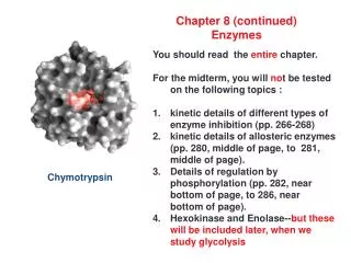
Chymotrypsin Mechanism Step 1: Substrate Binding Endo-peptidase specificity P3 - P2 - P1-|-P1’- P2’- P3’
Chymotrypsin MechanismStep 2: Nucleophilic Attack Serine protease – catalytic triad activates serine hydroxyl
Chymotrypsin MechanismStep 3: Substrate Cleavage N-terminal peptide released Covalent acyl intermediate
Peptidoglycan and Lysozyme • Peptidoglycan is a polysaccharide found in many bacterial cell walls • Cleavage of the cell wall leads to the lysis of bacteria • Lysozyme is an antibacterial enzyme
General Acid-Base + Covalent Catalysis: Cleavage of Peptidoglycan by Lysozyme X-ray structures of lysozyme with bound substrate analogs show that the C-1 carbon is located between Glu 35 and Asp 52 residues.