Gallstones: Types, Risk Factors, Management
1.59k likes | 2.19k Vues
Understand gallstones, types (cholesterol, pigment), risk factors (age, gender, diet), morphology, pathogenesis, natural history, and management options.

Gallstones: Types, Risk Factors, Management
E N D
Presentation Transcript
GALL STONES AND MANAGEMENT SANTHIP JOHN
GALL STONES • INTRODUCTION • TYPES • RISK FACTORS • MORPHOLOGY • PATHOGENESIS • NATURAL HISTORY • MANAGEMENT • SUMMARY • VIDEOCLIPS
GALL STONES • Most common biliary pathology affecting 10 to 20 % of adult population in developed countries. • Gallstones (choleliths) are crystalline bodies formed by accretion or concretion of normal or abnormal bile components.
Cholesterol stones 1. Pure [ 100 % ] 2. Mixed [> 70 %] Pigment stones [< 20 %] 1.Black stones 2.Brown stones Mainly two types
RISK FACTORS Classical risk factors– 5 F
1.Age-Prevalence increases with advancing age middle ageaffected more[ 40s] 2.Body habitus-Obesity, if you have high blood triglycerides,high fat diet 3.Childbearing -Pregnancy 4.Drugs - Fibric acid derivatives (or fibrates), oral contraceptives , postmenopausal estrogens, progesterone, octreotide ,ceftriaxone
5.Ethnicity - Pima tribe of Native Americans in Arizona&Scandinavians have an increased risk. Native Indian populations of Chile and Peru are highly susceptible, with a close to 100% lifetime risk of gall stones in their female population. 6.Family history - if your mother had gallstones 7.Gender - Females : Gallstones are found in 12% men and 24% women . 8.Hormones – Oestrogens
9.Impaired gall bladder emptying- such as truncal vagotomy, type 1 diabetes, octreotide, parenteral nutrition, and starvation/fasting or rapid voluntary weight loss Ileal and other metabolic diseases - [Crohn's disease, resection or bypass-where excess loss of bile salts occurs ] [Chronic hemolysis, alcoholic cirrhosis, biliary infection, primary biliary cirrhosis, duodenal diverticula, hyperparathyroidism]
CHOLESTEROL STONES • Higher consumption of dietary fat • Obesity • Hyperlipidemia syndromes • Advancing age • Female sex hormones • Rapid weight reduction • Inborn errors of bile acid metabolism
BLACK STONES • Chronic hemolytic syndromes • RBC destruction after cardiac valve replacement • Biliary infections • Ileal diseases like Crohn’s disease • Ileal ressection / Bypass • Cystic fibrosis with pancreatic insufficiency • Cirrhosis • Non-vegetarian food
BROWN STONES • Common in South East Asia • Ascaris ,E.coli ,Liver fluke [endemic]
Biliary stasis induced by worms leads to stone formation and then repeated bouts of cholangitis occurs- ORIENTAL CHOLANGIO HEPATITIS • Biliary stricture • Vegetarian food
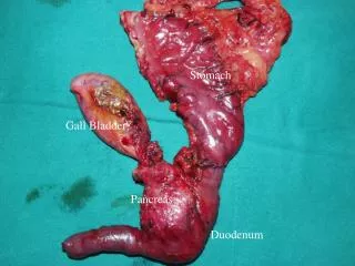
Stones can occur anywhere within the biliary tree. • In gall bladder : Cholelithiasis [Cholecystolithiasis] • In bile ducts : Choledocholithiasis • Stones may exist in both sites simultaneously also.
STONES IN GALL BLADDER[CHOLELITHIAIS] Can be Cholesterol or Black stones
Primary stones Stones which form de novo within bile ducts. [Brown stones] Secondary stones Stones which migrate from gall bladder to bile ducts. [Cholesterol stones] STONES IN BILE DUCT[CHOLEDOCHOLITHIAIS]
PRIMARYCHOLEDOCHOLITHIASIS -Arbitarily stones discovered more than two years after cholecystectomy . -It can occur in : • Post-traumatic biliary stricture • Narrowed biliary enteric anastomosis • Stenosis of sphincter Oddi • Sclerosing cholangitis and • Asian cholangiohepatitis
CHOLESTEROL STONES 3 STAGES 1.Supersaturation 2.Accelerated crystallisation 3.Formation of stones from crystals
BLACK STONES • Defective acidification of bile. • Increase in unconjugated bilirubin levels.
BROWN STONES • Precise mechanism- Unclear. • Bacterial contamination- [E. coli] –Beta glucuronidase- unconjugated bilirubin- then along with bacterial cell bodies form brown stones. • Vicious cycle continues.
PURE CHOLESTEROL STONE • 0.5 to 4 cm in diameter • Single • Ovoid/spherical in shape • Finely granular • Hard external surface • C/S :white to slight yellow with a more pigmented center – CHOLESTEROL SOLITAIRE
MIXED CHOLESTEROL STONE • 0.1 to 2 cm in diameter • Multiple • Mulberry/faceted shape • C/S : dark brown pigment is often found in the center of such stones
BLACK STONES • Usually less than 1cm in diameter • Jet black, brittle and often spiculated • They almost always form in gall bladder • Contains calcium bilirubinate,carbonate and phosphate
BROWN STONES • Usually less than 1cm in diameter • Brown or brownish yellow • Soft, soap like or greasy consistency • Often deformable • May form either in bile ducts or gall bladder • Contains calcium bilirubinate,palmitate and stearate
CHOLELITHIASISORCHOLEDOCHOLITHIASIS • 1. ASYMPTOMATIC • 2. SYMPTOMATIC • 3. COMPLICATED
SYMPTOMS OF CHOLELITHIASIS • BILIARY COLIC • DYSPEPTIC SYMPTOMS • OTHERS
Characteristics of pain • Episodic • Severe • Epigastrium/Right upper quadrant pain • Steady
SYMPTOMS OF CHOLEDOCHOLITHIASIS • BILIARY COLIC-Pain may be mild or severe and cannot be differentiated from pain arising in gall bladder. • NAUSEA & VOMITING-common • JAUNDICE-Intermittent/Progressive
Sometimes the stone may slip back into the body of gall bladder and the contents may escape by way of the cystic duct.This enables adequate drainage of the gall bladder and enables the pain to resolve.
COMPLICATIONS OF CHOLELITHIASIS Cholecystitis Gangrene Perforation Empyema Mucocoele Carcinoma OF CHOLEDOCHOLITHIASIS Cholangitis Acute pancreatitis Gall stone ileus
Continuous pain,Nausea,Vomiting,Pyrexia • O/E-Tenderness in (R) subcostal area Guarding, even rigidity [CHOLECYSTITIS] • MURPHY SIGN : Positive sign of cholecystitis. Painful arrest of respiration at the zenith of deep inspiration. Tenderness of the costal cartilage of 8th rib on the right side.
Sometimes gall bladder may : perforate localised peritonitis abscess[empyema]perforation septic peritonitis
MUCOCELE • Mucocele occurs when neck of gall bladder becomes obstructed by a stone but the contents remain sterile. • Bile is absorbed and replaced by gall bladder epithelium. • Gall bladder may be palpable.