Sensing and Acting
Sensing and Acting. Bats use sonar to detect their prey Moths, a common prey for bats, can detect the bat’s sonar and attempt to flee. Types of Sensory Receptors. Based on energy transduced, sensory receptors fall into five categories: Mechanoreceptors Chemoreceptors

Sensing and Acting
E N D
Presentation Transcript
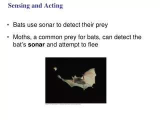
Sensing and Acting • Bats use sonar to detect their prey • Moths, a common prey for bats, can detect the bat’s sonar and attempt to flee
Types of Sensory Receptors • Based on energy transduced, sensory receptors fall into five categories: • Mechanoreceptors • Chemoreceptors • Electromagnetic receptors • Thermoreceptors • Pain receptors
Mechanoreceptors and Chemoreceptors • Mechanoreceptors sense physical deformation caused by stimuli such as pressure, stretch, motion, and sound • The sense of touch in mammals relies on mechanoreceptors that are dendrites of sensory neurons • General chemoreceptors transmit information about the total solute concentration of a solution
Cold Hair Pain Heat Light touch Epidermis Dermis Hypodermis Strong pressure Hair movement Connective tissue Nerve
Electromagnetic Receptors • Electromagnetic receptors detect electromagnetic energy, such as light, electricity, and magnetism • Some snakes have very sensitive infrared receptors that detect body heat of prey against a colder background
Eye Infrared receptor This rattlesnake and other pit vipers have a pair of infrared receptors, one between each eye and nostril. The organs are sensitive enough to detect the infrared radiation emitted by a warm mouse a meter away. Some migrating animals, such as these beluga whales, apparently sense Earth’s magnetic field and use the information, along with other cues, for orientation.
Thermoreceptors and Pain Receptors • Thermoreceptors, which respond to heat or cold, help regulate body temperature by signaling both surface and body core temperature • In humans, pain receptors, or nociceptors, are a class of naked dendrites in the epidermis • They respond to excess heat, pressure, or chemicals released from damaged or inflamed tissues
The mechanoreceptors involved with hearing and equilibrium detect settling particles or moving fluid • Hearing and perception of body equilibrium are related in most animals • Most invertebrates have sensory organs called statocysts • Statocysts contain mechanoreceptors and function in the sense of equilibrium • Many arthropods sense sounds with body hairs that vibrate or with localized “ears” consisting of a tympanic membrane and receptor cells
Ciliated receptor cells Cilia Statolith Sensory nerve fibers
Tympanic membrane 1 mm
Hearing and Equilibrium in Mammals • In most terrestrial vertebrates, sensory organs for hearing and equilibrium are closely associated in the ear • Vibrating objects create percussion waves in the air that cause the tympanic membrane to vibrate • The three bones of the middle ear transmit the vibrations to the oval window on the cochlea • These vibrations create pressure waves in the fluid in the cochlea that travel through the vestibular canal and strike the round window
Middle ear Outer ear Inner ear Semicircular canals Stapes Skull bones Middle ear Incus Auditory nerve, to brain Malleus Tympanic membrane Pinna Auditory canal Eustachian tube Cochlea Oval window Round window Tympanic membrane Eustachian tube Tectorial membrane Hair cells Bone Cochlea duct Auditory nerve Vestibular canal Tympanic canal Basilar membrane Axons of sensory neurons To auditory nerve Organ of Corti
Cochlea Stapes Axons of sensory neurons Vestibular canal Perilymph Oval window Apex Base Basilar membrane Tympanic canal Round window
Pressure waves in the canal cause the basilar membrane to vibrate, bending its hair cells • This bending of hair cells depolarizes their membranes, sending action potentials that travel via the auditory nerve to the brain
Semicircular canals Ampulla Flow of endolymph Flow of endolymph Vestibular nerve Cupula Hairs Hair cell Vestibule Nerve fibers Utricle Body movement Saccule
Hearing and Equilibrium in Other Vertebrates • Like other vertebrates, fishes and amphibians have inner ears near the brain • Most fishes and aquatic amphibians also have a lateral line system along both sides of their body • The lateral line system contains mechanoreceptors with hair cells that respond to water movement
Lateral line Lateral line canal Scale Opening of lateral line canal Neuromast Epidermis Lateral nerve Segmental muscles of body wall Cupula Sensory hairs Supporting cell Hair cell Nerve fiber
Smell in Humans • Olfactory receptor cells are neurons that line the upper portion of the nasal cavity • Binding of odorant molecules to receptors triggers a signal transduction pathway, sending action potentials to the brain
Brain potentials Action Olfactory bulb Nasal cavity Bone Odorant Epithelial cell Odorant receptors Chemoreceptor Plasma membrane Cilia Odorant Mucus
Structure of the Eye • Main parts of the vertebrate eye: • The sclera: white outer layer, including cornea • The choroid: pigmented layer • The conjunctiva: covers outer surface of sclera • The iris: regulates the pupil • The retina: contains photoreceptors • The lens: focuses light on the retina
Choroid Sclera Retina Ciliary body Fovea (center of visual field) Suspensory ligament Cornea Iris Optic nerve Pupil Aqueous humor Lens Central artery and vein of the retina Vitreous humor Optic disk (blind spot)
Humans and other mammals focus light by changing the shape of the lens • The human retina contains two types of photoreceptors: rods and cones • Rods are light-sensitive but don’t distinguish colors • Cones distinguish colors but are not as sensitive
Sensory Transduction in the Eye • Each rod or cone contains visual pigments consisting of a light-absorbing molecule called retinal bonded to a protein called opsin • Rods contain the pigment rhodopsin, which changes shape when absorbing light
Rod Outer segment Disks Inside of disk Cell body cis isomer Light Enzymes Synaptic terminal Cytosol Retinal Rhodopsin trans isomer Opsin
Hydrostatic Skeletons • A hydrostatic skeleton consists of fluid held under pressure in a closed body compartment • This is the main type of skeleton in most cnidarians, flatworms, nematodes, and annelids • Annelids use their hydrostatic skeleton for peristalsis, a type of movement on land produced by rhythmic waves of muscle contractions
Circular muscle contracted Circular muscle relaxed Longitudinal muscle relaxed (extended) Longitudinal muscle contracted Bristles Head
Exoskeletons and Endoskeletons • An exoskeleton is a hard encasement deposited on the surface of an animal • Exoskeletons are found in most molluscs and arthropods • An endoskeleton consists of hard supporting elements, such as bones, buried in soft tissue • Endoskeletons are found in sponges, echinoderms, and chordates
Key Head of humerus Axial skeleton Skull Examples of joints Appendicular skeleton Scapula Clavicle Shoulder girdle Scapula Sternum Ball-and-socket joints, where the humerus contacts the shoulder girdle and where the femur contacts the pelvic girdle, enable us to rotate our arms and legs and move them in several planes. Rib Humerus Vertebra Radius Humerus Ulna Pelvic girdle Carpals Ulna Phalanges Metacarpals Hinge joints, such as between the humerus and the head of the ulna, restrict movement to a single plane. Femur Patella Tibia Fibula Ulna Radius Pivot joints allow us to rotate our forearm at the elbow and to move our head from side to side. Tarsals Metatarsals Phalanges
Muscles move skeletal parts by contracting • The action of a muscle is always to contract • Skeletal muscles are attached in antagonistic pairs, with each member of the pair working against each other • A skeletal muscle consists of a bundle of long fibers running parallel to the length of the muscle • A muscle fiber is itself a bundle of smaller myofibrils arranged longitudinally
Human Grasshopper Extensor muscle relaxes Biceps contracts Tibia flexes Flexor muscle contracts Forearm flexes Triceps relaxes Biceps relaxes Extensor muscle contracts Tibia extends Forearm extends Flexor muscle relaxes Triceps contracts
The myofibrils are composed to two kinds of myofilaments: • Thin filaments consist of two strands of actin and one strand of regulatory protein • Thick filaments are staggered arrays of myosin molecules
Skeletal muscle is also called striated muscle because the regular arrangement of myofilaments creates a pattern of light and dark bands • Each unit is a sarcomere, bordered by Z lines • Areas that contain the myofilments are the I band, A band, and H zone
Muscle Bundle of muscle fibers Nuclei Single muscle fiber (cell) Plasma membrane Myofibril Light band Z line Dark band Sarcomere TEM 0.5 µm I band A band I band M line Thick filaments (myosin) Thin filaments (actin) Z line H zone Z line Sarcomere
The Sliding-Filament Model of Muscle Contraction • According to the sliding-filament model, filaments slide past each other longitudinally, producing more overlap between thin and thick filaments • As a result of sliding, the I band and H zone shrink
0.5 µm Z H I A Sarcomere Relaxed muscle fiber Contracting muscle fiber Fully contracted muscle fiber
The sliding of filaments is based on interaction between actin and myosin molecules of the thick and thin filaments • The “head” of a myosin molecule binds to an actin filament, forming a cross-bridge and pulling the thin filament toward the center of the sarcomere
Thick filament Thin filaments Thin filament Myosin head (low-energy configuration) Thick filament Cross-bridge binding site Thin filament moves toward center of sacomere. Actin Myosin head (low- energy configuration) Myosin head (high- energy configuration) Cross-bridge
The Role of Calcium and Regulatory Proteins • A skeletal muscle fiber contracts only when stimulated by a motor neuron • When a muscle is at rest, myosin-binding sites on the thin filament are blocked by the regulatory protein tropomyosin • For a muscle fiber to contract, myosin-binding sites must be uncovered • This occurs when calcium ions (Ca2+) bind to a set of regulatory proteins, the troponin complex
Tropomyosin Ca2+-binding sites Troponin complex Actin Myosin-binding sites blocked. Ca2+ Myosin- binding site Myosin-binding sites exposed.
Motor neuron axon Mitochondrion Synaptic terminal T tubule Sarcoplasmic reticulum Ca2+ released from sarcoplasmic reticulum Myofibril Sarcomere Plasma membrane of muscle fiber
The synaptic terminal of the motor neuron releases the neurotransmitter acetylcholine • Acetylcholine depolarizes the muscle, causing it to produce an action potential
Action potentials travel to the interior of the muscle fiber along transverse (T) tubules • The action potential along T tubules causes the sarcoplasmic reticulum to release Ca2+ • The Ca2+ binds to the troponin-tropomyosin complex on the thin filaments • This binding exposes myosin-binding sites and allows the cross-bridge cycle to proceed
Synaptic terminal of motor neuron PLASMA MEMBRANE Synaptic cleft T TUBULE ACh SR Ca2+ CYTOSOL Ca2+
Spinal cord Motor unit 2 Motor unit 1 Synaptic terminals Nerve Motor neuron cell body Motor neuron axon Muscle Muscle fibers Tendon
A motor unit consists of a single motor neuron and all the muscle fibers it controls • Recruitment of multiple motor neurons results in stronger contractions • Tetanus is a state of smooth and sustained contraction produced when motor neurons deliver a volley of action potentials
Types of Muscle Fibers • Skeletal muscle fibers are classified as slow oxidative, fast oxidative, and fast glycolytic • These categories are based on their contraction speed and major pathway for producing ATP • Cardiac muscle, found only in the heart, consists of striated cells electrically connected by intercalated discs • In smooth muscle, found mainly in walls of hollow organs, contractions are relatively slow and may be initiated by the muscles themselves
Animation and Videos • Bozeman - The Sensory System • Receptors in the Skin – 1 • Receptors in the Skin – 2 • The Human Eye • Vision • Information Processing in the Retina • Visual Patterns • Near and Distant Vision
Animation and Videos • Sound Transduction in the Human Ear • The Human Ear • Effect of Sound Waves on Cochlear Structures • Sound Transduction • Bozeman - The Skeletal System • Animation of Bone Growth • Osteoporosis • Balancing Calcium Levels in the Blood
Animation and Videos • Bozeman - The Muscular System • Action Potential and Muscle Contraction • Function of the Neuromuscular Junction • Motor Protein • Myofilament Contraction • Sarcomere Shortening • Breakdown of ATP and Cross-Bridge Movement During Muscle Contraction • Smooth Muscle Action