Download
1 / 3
30 likes | 35 Vues
The greater part of the tumors of the skull base may involve the sinuses or the ear or surrounding vascular structures. Modern Skull Base Surgery in India is carried out in association with ENT and Head Neck surgeon, Neurosurgeon, Vascular Surgeon, Interventional Radiologist.<br><br>

E N D
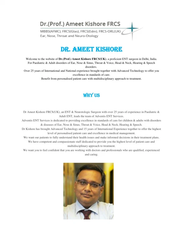
Skull Base Surgery In India - Dr. AmeetKishore What to expect from Skull Base Surgery inIndia? The greater part of the tumors of the skull base may involve the sinuses or the ear or surrounding vascular structures. Modern Skull Base Surgery in India is carried out in association with ENT and Head Neck surgeon, Neurosurgeon, Vascular Surgeon, InterventionalRadiologist. Endoscopic Pituitary tumourremoval This is a minimially invasive surgical procedure carried out through the nose and sinuses using endoscopes, to treat and remove tumours of the pituitary gland of the brain. This is a procedure carried out jointly with the the Neurosurgeon. An insignificantly intrusive method, called endoscopic endonasal surgery, utilizes a little entry point at the back of the nasal cavity and causes little disturbance of the nasal tissues. The ENT specialist works through the nostrils with a minor camera and light called an endoscope. In both systems, openings are made in the sphenoid sinus, and sella to reach the pituitary. Once the pituitary is uncovered, the neurosurgeon expels thetumor. Endoscopic CSF leakclosure If there is a hole in the bone and lining (dura) along the floor of the brain, CSF (cerebrospinal fluid) can leak into the nose. A minimally invasive proceure can be performed through the nose to repair this hole, stop the leak and thus prevent infection. Normally performed under general anesthesia, a specialist will work endoscopically through the nose to recognize the break and repair it utilizing muscle and other tissue from the body. CSF leaksurgery does not include cutting through the skin, as it is performed totally through the nostrils. Most patients should remain in the hospital for one to three days after surgery. A few patients may require a lumbar drain that is removed before goinghome.
Endoscopic Orbit and Optic nerve decopression An endoscopic procedure carreid out through the nose to decompress the eye ball or the optic (eye) nerve. This is sometimes required to reduce pressure on the eyeball or optic nerve and help treat certain types of visual loss. The objective of optic nerve decompression is to remove a bit of the hard optic channel, in this way removing pressure on the optic nerve. This method is finished with the utilization of endoscopes (little inflexible telescopes), enabling your specialist to experience the nose and sinuses to work in this sensitive surgery with no cuts to theface. Microscopic Facial nervedecompression The facial nerve that controls the muscles of the face travels through the ear bone. In certain types of facial paralysis, microscopic decompression of the facial nerve is performed to remove pressure on the nerve and allow recovery. By and large, facial nerve decompression is performed through the mastoid bone behind the ear. Bits of the bone are evacuated so that the compressed facial nerve can expandand the pressure that might bring about the loss offacial motionis removed. Contingent upon the individual case, the specialist may carry out the procedure through the middle cranial fossa technique or a mastoidectomy, orboth. Glomus tumourexcision These are slow growing benign vascular tumours of the ear and skull base. Of the various options available for their treatment, surgical removal may be one such. These operations are often carried out along with a Neurosurgeon. Fruitful removal of the tumor may likewise require delicateisolation of the tumor from the carotid supply route. This bit of the methodology is to a greatdegree
sensitive and requires the nearby collaboration of the head and neck specialist, ear specialist and, now and again, vascular andneurosurgeons. Acoustic Neuromaremoval These are slow growing benign tumours of the hearing and balance nerve. Of the various options available for their treatment, surgical removal may be one such. These operations are carried out along with a Neurosurgeon. A neurosurgeon or neuro-otologist can remove acoustic neuromas. The suboccipital approach is performed by a neurosurgeon and the translabyrinthine approach by the Neurotologist. Since every patient and every acoustic neuroma is unique, it is imperative to discuss the treatment that offers the full scope of alternatives, including surgery, radiation, and hearing and facial nerve preservation.