Download
1 / 14
140 likes | 358 Vues
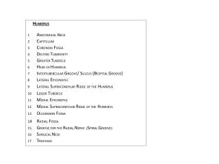
H UMERUS 1 A NATOMICAL N ECK 2 C APITULUM 3 C ORONOID F OSSA 4 D ELTOID T UBEROSITY 5 G REATER T UBERCLE 6 H EAD OF H UMERUS 7 I NTERTUBERCULAR G ROOVE / S ULCUS (B ICIPITAL G ROOVE ) 8 L ATERAL E PICONDYLE 9 L ATERAL S UPRACONDYLAR R IDGE OF THE H UMERUS

E N D
HUMERUS 1 ANATOMICALNECK 2 CAPITULUM 3 CORONOID FOSSA 4 DELTOID TUBEROSITY 5 GREATER TUBERCLE 6 HEAD OF HUMERUS 7 INTERTUBERCULAR GROOVE/ SULCUS (BICIPITAL GROOVE) 8 LATERAL EPICONDYLE 9 LATERALSUPRACONDYLARRIDGEOF THEHUMERUS 10 LESSERTUBERCLE 11 MEDIAL EPICONDYLE 12 MEDIALSUPRACONDYLARRIDGEOF THEHUMERUS 13 OLECRANONFOSSA RADIALFOSSA GROOVEFOR THERADIALNERVE (SPIRALGROOVE) 16 SURGICAL NECK 17 TROCHLEA
ULNA 1 BODY OF THE ULNA 2 CORNOIDPROCESS 3 HEAD OF ULNA 4 INTEROSSEUSBORDER OF THE ULNA 5 OLECRANON PROCESS 6 RADIAL NOTCH OF THE ULNA 7 STYLOID PROCESS OF THE ULNA 8 TROCHLEAR NOTCH OF THE ULNA • 9 ULNARTUBEROSITY(TUBEROSITY OF THE ULNA) RADIUS 1 BODY OF THE RADIUS 2 DORSAL TUBERCLE 3 HEAD OF THE RADIUS 4 INTEROSSEUS BORDER OF THE RADIUS 5 INTEROSSEUS MEMBRANE OF THE FOREARM 6 NECK OF THE RADIUS 7 RADIAL TUBEROSITY (TUBEROSITY OF THE RADIUS) 8 STYLOIDPROCESS OF THE RADIUS 9 ULANAR NOTCH OF THE RADIUS
View? • View? • 6 • 1 • 5 • 7 • 4 • 9
View? • View? • 10 • 10 • 12 • 8 • 2 • 11 • 3 • 11 • 17 • 17
View? • 14 • 9 • 4 • 5 • 8 • 3
Lateral of Right Humerus • 2 • 3 • 7 • 6 • 6 • 1 • 1 • 4 • 3 • 2 • 7 • 9
16 • 5 • 8 ON PROCESS • 2 • 9 • 15 • 13
Lateral of Right Humerus • Lateral of Right Humerus
Medial of Right Humerus • Anterior of Right Humerus
The epiphyseal plate (or growth plate) is a hyaline cartilage plate in the metaphysis at each end of a long bone. The plate is found in children and adolescents; in adults, who have stopped growing, the plate is replaced by an epiphyseal line. Whereas endochondral ossification is responsible for the initial bone development from cartilage in utero and infants, the epiphyseal plate is responsible for longitudinal growth of bones. The plate's chondrocytes are under constant division by mitosis. These daughter cells stack facing the epiphysis while the older cells are pushed towards the diaphysis. As the older chondrocytes degenerate, osteoblasts ossify the remains to form new bone. Around the end of puberty, the epiphyseal cartilage cells stop duplicating and the entire cartilage is slowly replaced by bone, leaving only a thin epiphyseal line.[1] Once the adult stage is reached, the only way to manipulate height is modifying bone length via distraction osteogenesis. The growth plate has a very specific morphology in having a zonal arrangement. The growth plate includes a relatively inactive reserve zone at the epiphyseal end, moving distally into a proliferative and then hyper trophic zone and ending with a band of ossifying cartilage (the metaphysis). The growth plate is clinically relevant in that it is often the primary site for infection, metastasis, fractures and the effects of endocrine bone disorders