Adrenal Glands
Adrenal Glands. Learning objectives: The student should: Recognize the variants of hyperadrenalism Recognize the variants of hypoadrenalism

Adrenal Glands
E N D
Presentation Transcript
Adrenal Glands • Learning objectives: • The student should: • Recognize the variants of hyperadrenalism • Recognize the variants of hypoadrenalism • Understand the clinical overview, the histopathological features and molecular pathology of both medullary (pheochromocytoma) and adrenocorticalneoplasms .
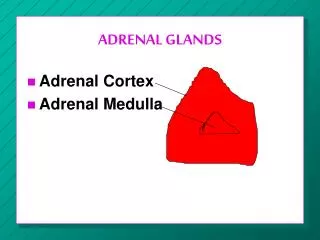
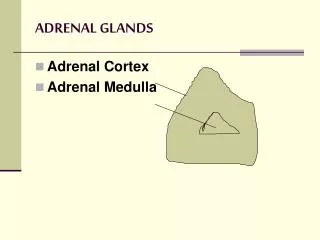
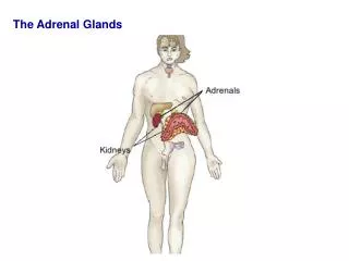
Adrenal Glands • The adrenal glands: paired endocrine organs: cortex and medulla: 4 layers • Three layers in the cortex: • Zonaglomerulosa • Zonareticularis abuts the medulla. • Intervening is the broad zonafasciculata (75%) of the total cortex.
Adrenal Gland Three types of steroids: (1) Glucocorticoids (principally cortisol) zonafasciculata (2) Mineralocorticoids (aldosterone) zonaglomerulosa (3) Sex steroids (estrogens and androgens) zonareticularis. • The adrenal medulla chromaffin cells- catecholamines, mainly epinephrine
ADRENOCORTICAL HYPERFUNCTION • Three basic types of corticosteroids (glucocorticoids, mineralocorticoids, and sex steroids) • Three distinctive hyperadrenal syndromes: (1) Cushing syndrome, characterized by increased cortisol (2) Hyperaldosteronism (3) Adrenogenital or virilizing syndromes caused by an excess of androgens
Hypercortisolism (Cushing Syndrome) • Broadly divided into exogenous and endogenous causes. • The vast majority of cases of Cushing syndrome are the result of the administration of exogenous glucocorticoids (“iatrogenic” Cushing syndrome). • The endogenous causes can, in turn, be divided into those that are ACTH dependent and those that are ACTH independent
ADRENOCORTICAL HYPERFUNCTION, Morphology One of the following abnormalities: • Cortical atrophy: results from exogenous glucocorticoids • Diffuse hyperplasia: individuals with ACTH-dependent Cushing’s syndrome • Macronodular (less than 3cm), or micronodular(1-3mm) hyperplasia • Adenoma or carcinoma
Hyperaldosteronism Excess aldosterone secretion • Primary aldosteronism (autonomous overproduction of aldosterone) with resultant suppression of the renin-angiotensin system and decreased plasma renin activity • Secondaryhyperaldosteronism, in contrast, aldosterone release occurs in response to activation of the renin-angiotensin system
Hyperaldosteronism, Clinical • Presents with hypertension. With an estimated prevalence rate of 5% to 10% among non-selected hypertensive patients. • Primary hyperaldosteronism may be the most common cause of secondary hypertension (i.e., hypertension secondary to an identifiable cause). • Aldosterone promotes sodium reabsorption. • Hypokalemia results from renal potassium wasting
Aldosterone-producing adenomas , Morphology • Solitary • Small (<2 cm in diameter), bright yellow on cut section • Well-circumscribed lesions left > right • Thirties and forties • Women more often than in men • Buried within the gland and do not produce visible enlargement • Bright yellow on cut section
Adrenocortical Insufficiency • Caused by either primary adrenal disease or decreased stimulation of the adrenals due to a deficiency of ACTH (secondary hypoadrenalism)
Adrenocortical Insufficiency • Three patterns of adrenocortical insufficiency (1) Primary acuteadrenocortical insufficiency (adrenal crisis) (2) Primary chronic adrenocortical insufficiency (Addison disease), and (3) Secondary adrenocortical insufficiency
Pheochromocytoma • Pheochromocytomas(chromaffin cells ) catecholamines • Similar to aldosterone-secreting adenomas, give rise to surgically correctable forms of hypertension. • 0.1% to 0.3%( fatal ) • Other peptides –Cushing etc…
Pheochromocytoma "rule of 10s": • 10% of pheochromocytomas arise in association with one of several familial syndromes MEN-2A and MEN-2B syndromes. • 10% of pheochromocytomas are extra-adrenal. • 10% of nonfamilial adrenal pheochromocytomas are bilateral; this figure may rise to 70% in cases that are associated with familial syndromes. • 10% of adrenal pheochromocytomas are biologically malignant • 10% of adrenal pheochromocytomas in childhood
Pheochromocytoma Syndrome Components MEN, type 2A :Medullary thyroid carcinomas and C-cell hyperplasia, Pheochromocytomas and adrenal medullary hyperplasia, Parathyroid hyperplasia MEN, type 2B : Medullary thyroid carcinomas and C-cell hyperplasia, Pheochromocytomas and adrenal medullary hyperplasia, Mucosal neuromas, Marfanoid features
Pheochromocytoma Von Hippel-Lindau:Renal, hepatic, pancreatic, and epididymal cysts, Renal cell carcinomas, Angiomatosis, Cerebellar hemangioblastomas, DM Von Recklinghausen Neurofibromatosis :Café au lait skin spots, Schwannomas, meningiomas, gliomas Sturge-Weber: Cavernous hemangiomas of fifth cranial nerve distribution
Pheochromocytoma • Small to large hemorrhagic • Well demarcated • Polygonal to spindle shaped (chromaffin, chief cells) • Sustentacular small cells • Together, Zellballen nests