Download
1 / 32
410 likes | 2.68k Vues
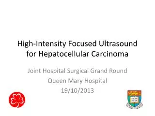
High Intensity Focused Ultrasound (HIFU) guided by MRI. Christakis Damianou Frederick Institute of Technology, MEDSONIC LTD November 9, 2006 HIFU Basics. Diagnostic ultrasound intensities: < 1 W/cm 2 HIFU intensities: 500-10000 W/cm 2 . Friction with tissue causes heating.

E N D
High Intensity Focused Ultrasound (HIFU) guided by MRI. Christakis Damianou Frederick Institute of Technology, MEDSONIC LTD November 9, 2006
HIFU Basics. • Diagnostic ultrasound intensities: < 1 W/cm2 • HIFU intensities: 500-10000 W/cm2. • Friction with tissue causes heating. • Temperature produced is in the range of 60-100 oC. • Tumors cells are destroyed due to excessive heating. • Spherically focused ultrasound produces localized heating.
MRI/HIFU System Damianou C. et al., Journal of Ultrasound in Medicine and Biology Vol. 30 (3), pp. 397-404, 2004
Video of lesion development in kidney and muscle. • Pulse duration= 10 s
MR compatible transducer. MR compatible transducer Non-MR compatible transducer
MRI monitoring of Ultrasound Hynynen K., Damianou C. “Utrasound surgery", Ultrasound Med. Biol., 1992;19 (1): 91-97.
T2-W FSE: effect of TE. TE=32 ms TE=64 ms C. Damianou et al. Journal of Ultrasound in Medicine & Biology 2004:30 (3):397-404.
Effect of TR on T1-Weighted FSE. TR=300 ms TR=400 ms TR=500 ms TR=700 ms
T1W FSPGR: effect of TR. 2000 W/cm2 for 5 s TR=100 ms TR=50 ms (4x4 lesions) 1 lesion created using cavitation. TR=150 ms TR=300 ms
MRI Monitoring of HIFU in liver. T2-FSE T1-weighted FSE Proton Density Photo
MRI Monitoring of HIFU in liver. MRI(Proton density) Photograph Pathology
Cavitation detection. T1W FSE T2W FSE FSPGR
Cavitation detection. T1W FSE T2W FSE T1W FSPGR
T2W FSE-Comparison of thermal vs. cavitation. 1000 W/cm2 (30 s) 3500 W/cm2 (30 s)
Temperature elevation-thermal mode. Each MRI image is acquired in 5 s (FSPGR). 1000 W/cm2 for 5 s
In vivo results (T2W FSE). 1000 W/cm2 2000 W/cm2 3500 W/cm2
Complete coverage Incomplete coverage C. Damianou, Journal of Ultrasound in Medicine and Biology 2004; 30 (9):1209-1215.
Brain tumor strategy. Astrocytoma
MR compatible robot. PCT/CY2006/000001, 19/1/2006
Brain ablation under MRI guidance. T1 FSE T2 FSE Work done by: K. Ioannides FLAIR
Brain ablation. FLAIR T1-FSE C. Damianou, K. Ioannides, J of Ultrasound in Medicine and Biology, in press.
Brain ablation. T1 FSE TR=300 ms TR=500 ms
Brain ablation. FLAIR
Acknowledgments • K. Ioannides (M.D.), M. Komodormos (Ph.D.), Y. Chapelon (Ph.D.), N. Milonas (M.Sc.) Work supported by • ULTRASOUND I, (Res. Prom. Found.) • SONOTHERM (Res. Prom. Found.) • ULTRASOUND II (Res. Prom. Found.) • TROY (FP6) • N2L (FP6) • CDTU (Medical Research foundation of France) • MARiUS (FP6) • SONOMRI (Ministry of industry of Cyprus)