
The Endocrine System
E N D
Presentation Transcript
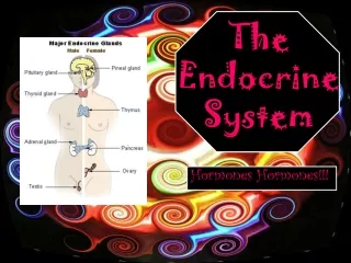
1. The Endocrine System
2. Cell Communication � 4 ways
Gap Junctions
Neurotransmitters
Paracrines
Hormones
3. Comparison of the Nervous System and the Endocrine System
5. Similarities Between Neurotransmitters and Hormones
Several chemicals function as both neurotransmitters and hormones � norepinephrine, dopamine, antidiuretic hormone
Neuroendocrine cells � neurons that release their secretions into the extracellular fluid � oxytocin and the catecholamines
Overlapping effects � Norepinephrine and glucagon cause glycogen hydrolysis
9. The Hypothalamus and Pituitary Gland
Pituitary gland is suspended from the hypothalamus by a stalk called the infundibulum � housed in the sella turcica of the sphenoid bone
Grows 50% in pregnancy
Composed of two structures � the adenohypohysis and the neurohypohysis
Adenohypophysis arises from a hypophyseal pouch that grows upward from the pharynx
Neurohypophysis arises from the neurohypophyseal bud which is a downgrowth of the brain
10. Adenohypophysis constitutes the anterior three fourths of the pituitary and is generally referred to as the anteror pituitary
Has no nervous connection to the hypothalamus
Connected to the hypothalamus by a complex of vessels called the hypophyseal portal system � begins with primary capillaries in the hypothalamus which lead to portal venules that lead down the pituitary stalk to a complex of secondary capillaries in the anterior pituitary
11. The primary capillaries pick up hormones from the hypothalamus , the venules deliver them to the anterior pituitary and the hormones leave the circulation at the secondary capillaries
The pituitary has especially porous capillaries called fenestrated capillaries that allow for the uptake and release of hormones
12. Neurohypophysis constitutes the posterior one-fourth of the pituitary and is referred to as the posterior pituitary
Has a median eminence and a stalk
The neurohypophysis is a mass of neuroglia and nerve fibers
The nerve fibers arise from cell bodies in the hypthalamus, travel down the stalk as a bundle called the hypothalamo-hypophyseal tract and end in the posterior lobe
The hypothalamic neurons synthesize hormones, transport them down the tract, and store them in the posterior pituitary until a nerve signal triggers their release
19. Hypothalamic Hormones
Hypothalamus produces eight hormones of interest to this course � six of these travel through the portal system and regulate the anterior pituitary � four of these are releasing hormones and two are inhibiting hormones
Two other hypothalamic hormones are secreted by way of the posterior pituitary �oxytocin is produced by neurons in the paraventricular nucleus of the hypothalamus and Antidiuretic hormone is produced neurons in the supraoptic nucleus of the pituitary
21. Pituitary Hormones � anterior lobe secretes six main hormones and the first five of these are trophic hormones that stimulate endocrine cells elsewhere to release their own hormones
FSH and LH are called gonadotropins
Three Axes � hypothalamo-pituitary-gonadal axis - hypothalamo-pituitary-thyroid axis � hypothalamo-pituitary-adrenal axis
The posterior pituitary produces no hormones of its own, but stores and releases OT and ADH
23. Actions of the Pituitary Hormones
Follicle Stimulating Hormone � In the ovary it stimulates development of eggs and follicles and in the testes it stimulates sperm production
Lutenizing Hormone � in the female it stimulates ovulation and stimulates the corpus luteum to secrete estrogen and progesterone � in the male it stimulates the interstitial cells of the testes to secrete testosterone
24. Thyroid stimulating hormone (TSH) � stimulates growth of the thyroid gland and the secretion of thyroid hormone
Adrenotrophic hormone (ACTH) � stimulates the adrenal cortex to secrete its hormones (corticosteroids) especially cortisol
Prolactin (PRL) � stimulates the mammary glands to produce milk � in the male it enhances the secretion of testosterone
Growth Hormone (GH) � stimulates the liver to produce insulin like growth factors (IGF-II, II
25. GH- IGF affects protein synthesis, lipid metabolism, carbohydrate metabolism, electrolyte balance, bone growth, cartilage growth, and muscle growth
26. Antidiuretic hormone (ADH) � acts on the kidney to increase water retention, reduce urine volume, prevent dehydration, causes vasoconstriction, acts as a brain neurotransmitter
Oxytocin (OT)- stimulates smooth muscle of the uterus thus stimulates labor, stimulates the flow of milk in the mammary gland
28. Negative Feedback Inhibition
The pituitary stimulates another endocrine gland to secrete its hormone and that hormone feeds back to the pituitary and inhibits that trophic hormone.
Negative feedback in the hypothalamic- pituitary-thyroid axis is as follows
The hypothalamus secretes thyrotropin releasing hormone (TRH)
TRH stimulates the anterior pituitary to secrete thyroid stimulating hormone (TSH)
29. TSH stimulates the thyroid gland to secrete T3 and T4
T3 and T4 stimulate the metabolism of most cells throughout the body
T3 and T4 also inhibit the release of TSH by the pituitary
T3 and T4 inhibit the release of TRH by the hypothalamus
This process maintains homeostasis
Feedback is not always negative
31. Pineal Gland
Found in children and involutes to a small mass of fibrous tissue in the adult
Secrete seratonin and melatonin - the significance of which is uncertain
There seems to be a relationship between melatonin and mood disorders
Pineal tumors can cause premature onset of puberty in boys
32. Thymus
Located in the mediastinum
Large in infants and involutes after puberty
Affects the development of disease fighting T lymphocytes
33. Thyroid Gland
The largest endocrine gland
It arises from the root of the embryonic tongue and descends into the neck
After birth it is wrapped around the anterior and lateral aspects of the trachea immediately below the larynx
It consists of two large lobes, one on each side of the trachea, connected by an anterior isthmus
34. Histologically composed of thyroid follicles � each filled with a protein rich colloid � lined by a simple cuboidal epithelium of follicular cells
Follicular cells secrete T3 or triiodothyronine and T4 or thyroxine � the expression, thyroid hormone, refers to T3 and T4 collectively
Thyroid hormone is secreted in response to TSH
Increases body�s metabolic rate
Raises oxygen consumption
Increases heat production
35. TH secretion rises in cold weather to help compensate for heat loss
Raises the heart rate and contraction strength
Raises the respiratory rate
Stimulates the appetite and accelerates the breakdown of carbohydrates, fats and protein
Promotes alertness
Promotes bone growth and remodeling
Involved in the development of the skin, hair, teeth, and nails
Involved in the development of the fetal nervous system and the skeleton
36. The thyroid also secretes calcitonin which lowers the serum calcium levels by decreasing osteoclastic activity and increasing osteoblastic activity
Calcitonin is secreted by C cells which are located between the thyroid follicles
42. Parathyroid Glands
Partially embedded in the posterior surface of the thyroid
Usually two on each side
Secrete Parathyroid hormone (PTH) in response to hypocalcemia
PTH promotes synthesis of calcitriol which promotes calcium absorption in the intestine
PTH inhibits urinary calcium excretion
PTH stimulates osteoclastic activity
In low doses PTH can stimulate osteoblastic activity
44. Adrenal Medulla
Consists of modified neurons, chromaffin cells, that lack dendrites and axons
These cells are innervated by sympathetic preganglionic fibers
The chromaffin cells secrete epineprine (85%) and norepinephrine
These hormones supplement the effects of the sympathetic nervous system � increase alertness
Epinephrine boosts glucose levels by promoting glycogenolysis and gluconeogenesis (conversion of fatty acids, amino acids to glucose)
45. Epinephrine inhibits insulin secretion � thus it is glucose sparing
The catecholamines increase heart rate and blood pressure, increase pulmonary airflow, raise the metabolic rate, and inhibit urination and digestion
46. Adrenal Cortex � three areas � zona glomerulosa � zona fasciculata � zona reticularis
The cortex synthesizes more than 25 steroid hormones known collectively as corticosteroids
Zona glomerulosa secretes mineralocoticoids the main one being aldosterone � aldosterone promotes sodium retention and potassium excretion by the kidney
47. The Zona fasciculata secretes glucocorticoids with the main one being cortisol
Glucocorticoids stimulate fat and protein catabolism, promote gluconeogenesis � they have an anti-inflammatory effect and long term secretion suppresses the immune system
48. The Zona reticularis secretes androgens and estrogens
Androgens control many aspects of male development � the main androgen is dehydroepiandrosterone (DHEA) - converted to a more potent androgen, testosterone
This source is relatively unimportant in men because the testes secretes most of the testosterone � in females the adrenal secretes 50% of the total androgen requirement
In both sexes androgens stimulate the growith of axillary and pubic hair and sustain libido throughout adult life
49. Adrenal estrogen is a relatively unimportant source of estrogen as most of this come from the ovary � after menopause the adrenal becomes the estrogen source for the female
Both estrogens and androgens promote adolescent skeletal growth and help maintain adult bone mass
52. Pancreas � located inferior and dorsal to the stomach � mostly an exocrine digestive gland � scattered through the exocrine tissue are endocrine cell clusters called Islets of Langerhans � they constitute 2% of the pancreatic tisssue � they secrete three important hormones
Insulin � secreted by beta cells � stimulates cells to absorb glucose and amino acids from the blood - stimulates muscle and adipose to store glycogen and fat � suppresses the use of already stored fuels � promotes glycogen, fat and protein synthesis � antagonizes the effects of glucagon � kidney, brain, liver and red blood cells do not depend on insulin for glucose uptake although it does promote glycogen synthesis in the liver
53. Glucagon � secreted by alpha cells when blood glucose levels fall between meals �
In the liver it stimulates gluconeogenesis, glycogenlolysis and the release of glucose into the circulation
In the adipose tissue it stimulates fat catabolism and the release of free fatty acids
Glucagon is also secreted in response to rising amino acid levels in the blood and promotes amino acid absorption
54. Somatostatin � secreted by delta cells � inhibits the secretion of insulin and glucagon by the beta and alpha cells � reason for this is unclear
57. The Gonads � both endocrine and exocrine � their exocrine products are eggs and sperm and their endocrine products are the gonadal hormones, most of which are steroids
Each follicle of the ovary contains an egg cell surrounded by granulosa cells which produce estradiol during the first half of the menstrual cycle � after ovulation the corpus luteum secretes estradiol and progesterone for12-14 days and for 10-12 weeks in the event of a pregnancy.
58. The sex hormones contribute to the development of the reproductive system and feminine physique, promote adolescent bone growth, regulate the menstrual cycle, sustain pregnancy, and prepare the mammary glands for lactation
The follicle and corpus luteum also secrete inhibin which suppresses FSH secretion
59. The testis � consists of seminiferous tubules that produce sperm � between them are clusters of interstitial cells which produce testosterone
Testosterone stimulates development of the male reproductive system in the fetus and the adolescent , the development of the masculine physique, and sex drive � it sustains sperm production
Sertoli cells of the testis secrete inhibin which supresses FSH and stablizes the rate of sperm production
65. Hormone chemistry
Steroid hormones � derived from cholesterol-have a four ring backbone � includes sex steroids produced by the ovaries and testis, and the corticosteroids in the adrenal cortex
Peptide hormones � chains of 3-200 or more amino acids � the two posterior pituitary hormones OT and ADH are very similar oligopeptides � except for dopamine the releasing and inhibiting hormones produced by the hypothalamus are peptides
66. Most hormones of the anterior pituitary are polypeptides or glycoproteins � all glycoproteins have anidentical alpha chain of 92 amino acids and a variable beta chain that distinguishes them from each other
Monoamines � made from amino acids and have an amino group
72. Hormone Synthesis
Steroids � synthesized from cholesterol � differ mainly in the functional groups attached to the four-ringed steroid backbone
74. Peptides � synthesized the same way as any other protein � the gene for the hormone is transcribed to form a molecule of mRNA � ribosomes translate the mRNA and assemble amino acids in the right order � the newly synthesized hormone is a pre prohormone � signal peptide guides it to the rough endoplamic reticulum where the signal peptide is split off and now there is a prohormone � the prohormone is transferred to the Golgi apparatus where it is packaged for secretion
76. Monoamines - Thyroid hormone synthesis
Follicular cells absorb I ions from the blood plasma and secrete them into the lumen of the follicle � here the I ion is oxidized to a neutral I atom
Meanwhile the follicular cells synthesize a large protein called thyroglobulin through the usual mechanism involving the rough endoplasmic reticulum and Golgi apparatus � this is released into the lumen by exocytosis � each thyroglobulin molecule has 123 tyrosines
77. In the lumen tyrosine and iodine combine to form T3 and T4 � the TH remains bound to the thyroglobulin in storage in the follicle until the cell receives a signal (TSH) inducing it to secreteTH
When stimulated by TSH the follicular cells absorb thyroglobulin by pinocytosis and liberates T3 and T4 from it
The two hormones are released into the blood a s 10% T3 and 90% T4 � they bind to various transport proteins that carry them to their target cells
80. Hormone Transport
Steroids and thyroid hormone are hydrophobic and require hydrophilic transport proteins to get to their target
Transport proteins are albumins and globulins and are synthesized by the liver
Hormones can be bound or unbound but only unbound hormones can leave the blood stream and get to a target cell
Transport proteins also protect hormones from being broken down by enzymes and prolong their half life
81. Thyroid hormone binds to three transport hormones - albumin, thyretin, and thyroxine binding globulin (TBG)
TBG binds 99.8% of T3, 99.98% of T4 � bound TH serves as a reservoir � if thyroid is surgically removed there is enough TH for about 2 weeks
Steroid hormones bind to globulins such as transcortin which binds cortisol
Aldosterone has no specific transport protein and therefore has a very short half life
Sex hormones are bound to SHBG
82. Hormone Receptors - hormones stimulate only those cells that have receptors for them
The receptors are protein molecules located on the plasma membrane, on mitochondria, on other organelles or on the nucleus
Specificity means that the receptor for one hormone will not bind other hormones
Saturation is the condition in which all receptor hormones are occupied by hormone molecules
83. Steroids and Thyroid hormones and Their Receptors
These hydrophobic hormones easily penetrate the phospholipid plasma membrane
Steroids enter the nucleus and bind to a receptor that has three functional areas � one that binds the hormone, one that binds to an acceptor site on the chromatin and one that activates DNA transcription
Transcription produces new mRNA that leads to the synthesis of proteins that alter the metabolism of the target cell � estrogen leading to the development of progesterone receptors in the endometrium is an example and this is followed by progesterone receptors and gylgogen
84. Thyroid Hormone receptors
Unbound T3 and T4 enter the target cell cytoplasm where T4 is converted to T3 �
T3 binds to receptors in three sites � on mitochondria where it increases the rate of aerobic respiration, in the nucleus where it stimulates DNA transcription and mRNA synthesis , and on the ribosomes where it stimulates translation of mRNA and thereby increases the rate of protein synthesis
One of the proteins synthesized is Na- K ATPase which is the sodium �potassium pump � this generates heat
87. Hormone Receptors for Peptides and Catecholamines
Peptides and chatecolamines are hydrophylic and cannot penetrate the cell wall of the target cell � they bind to a cell-surface receptor which is linked to a second messenger system
Cyclic AMP second messenger system is seen with ACTH, FSH, LH, PTH, TSH, Glucagon, Calcitonin, and Catecholamines
89. Diacylglycerol (DAG) second messenger and Inositol triphoaphate (IP3)pathways � seen with ADH, TRH, OT, LHRH, Catecholamines
OT works through the IP3 second messenger system which raises calcium levels which stimulate the smooth muscle of the uterus
91. Eicosanoids and Paracrine Signaling
Paracrine messengers are chemical signals released by cells into the tissue fluid � they do not travel to their target cells by way of the blood � they diffuse to nearby cells � examples are histamine, nitric oxide, and some catocholamines
Eicosanoids are a family of paracrine secretions
Arachidonic acid is liberated from the phospholipids of the plasma membrane by the action of phospholipase A2
92. Lipoxygenase converts arachidonic acid to leukotrienes
Leukotrienes promote vasoconstriction, bronchospasm, and increased blood vessel permeability
Cyclooxygenase converts arachidonic acid to thromboxanes which stimulate vasoconstriction and clotting, prostacyclins which inhibit blood clotting and vasoconstriction and prostaglandins which have several function
95. Endocrine Disorders
Diabetes insipidus
Toxic goiter ( Grave�s disease)
Hypothyroidism � myxedema
Hypertyroidism
Endemic goiter
Hyperparathyroidism
Hypoparathyroidism
Cushing�s syndrome
Addison disease
96. Adrenogenital syndrome
Pheochromocytoma
Acromegally
Gigantism
Pituitary dwarfism
Androgen insensitivity syndrome
Diabetes Mellitus � types I and II