Download

1 / 27
320 likes | 775 Vues
Flagella and Cilia. A. P. Biology Chapter 6 Mr. Knowles Liberty Senior High School. Flagella of Prokaryotes (Bacteria). Composed of a flagellin subunit. Usually sheathed (covered). Rotates by way of a basal body in the bacterial cell. Unique to bacteria.

E N D
Flagella and Cilia A. P. Biology Chapter 6 Mr. Knowles Liberty Senior High School
Flagella of Prokaryotes (Bacteria) • Composed of a flagellin subunit. • Usually sheathed (covered). • Rotates by way of a basal body in the bacterial cell. • Unique to bacteria.
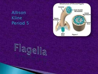
Eukaryotic Flagella • Completely different than bacteria. • Circle of 9 fused pairs of microtubules that make a cyclinder. • 2 unfused microtubules in the center of cylinder. • Called the 9 + 2 structure.
Eukaryotic Flagella • Whip-like appendage, used in movement and longer than cilia. • Is an outward projection of cytoplasm.
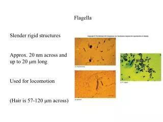
Direction of swimming (a) Motion of flagella. A flagellum usually undulates, its snakelike motion driving a cell in the same direction as the axis of the flagellum. Propulsion of a human sperm cell is an example of flagellatelocomotion (LM). 1 µm Flagella beating pattern Figure 6.23 A
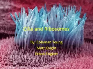
Cilia • More numerous than flagella. • Cilia of unicellular eukaryotes = movement of cell. Ex. Paramecium
(b) Motion of cilia. Cilia have a back- and-forth motion that moves the cell in a direction perpendicular to the axis of the cilium. A dense nap of cilia, beating at a rate of about 40 to 60 strokes a second, covers this Colpidium, a freshwater protozoan (SEM). Figure 6.23 B Ciliary Motion 15 µm
Cilia • Cilia of multicellular eukaryotes = movement of debris, sensory cells of vertebrate ear, epithelia of respiratory and reproductive tracts. • Have similar microtubule structure of 9 + 2 as eukaryotic flagella.
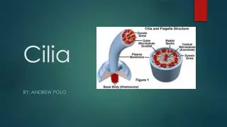
Outer microtubule doublet Plasma membrane 0.1 µm Dynein arms Central microtubule Outer doublets cross-linking proteins inside Microtubules Radial spoke Plasma membrane Basal body (b) 0.5 µm 0.1 µm (a) Triplet (c) Figure 6.24 A-C Cross section of basal body Cilia and flagella share a common ultrastructure
Microtubule doublets ATP Dynein arm (a) Powered by ATP, the dynein arms of one microtubule doublet grip the adjacent doublet, push it up, release, and then grip again. If the two microtubule doublets were not attached, they would slide relative to each other. Figure 6.25 A Protein Dynein: • Is responsible for the bending movement of cilia and flagella
ATP Outer doublets cross-linking proteins Anchorage in cell (b) In a cilium or flagellum, two adjacent doublets cannot slide far because they are physically restrained by proteins, so they bend. (Only two of the nine outer doublets in Figure 6.24b are shown here.) Figure 6.25 B
1 3 2 (c) Localized, synchronized activation of many dynein arms probably causes a bend to begin at the base of the Cilium or flagellum and move outward toward the tip. Many successive bends, such as the ones shown here to the left and right, result in a wavelike motion. In this diagram, the two central microtubules and the cross-linking proteins are not shown. Figure 6.25 C
Microvillus Plasma membrane Actin Filaments Intermediate filaments 0.25 µm Figure 6.26 • Are found in microvilli