Download

1 / 10
130 likes | 533 Vues
Cilia and Flagella. Structure and Function in Eukaryotes. By Justin Robbins and Katrina Truebenbach. Overview. Cilia and Flagella are organelles that are primarily used for the transportation of the cell. They propel the cell by flicking back and forth.

E N D
Cilia and Flagella Structure and Function in Eukaryotes By Justin Robbins and Katrina Truebenbach
Overview • Cilia and Flagella are organelles that are primarily used for the transportation of the cell. They propel the cell by flicking back and forth. • Cilia are short and reminiscent of hairs. There are many per cell. • Flagella are longer and there are far fewer per cell. They are reminiscent of a tail.
Real-life Examples:Eukaryotes • Most common in single-celled organisms (protists). • However, some multi-cellular organisms have cilia and flagella. • Human windpipe cells and some lung cells have cilia to clean the respiratory system of breathing hazards. • Fish have cilia to help bring water through the gills. • Many types of sperm have flagella to help them move.
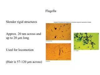
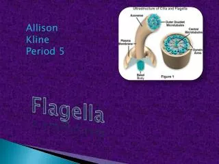
Structure: 9+2 Pattern • Cilia or flagella is composed of microtubules that are encased in a plasma membrane. This bundle of microtubules is called the axoneme. • A plasma membrane is made of lipids and proteins and is essentially the same as a normal cell membrane. • There are 9 pairs of connected microtubules in a circle towards the outside edge of the cilia/flagella. These are called the outer microtubule doublets. • The outer microtubules are connected to each other in a ring with cross-links (not pictured). • The outer microtubules also connect to the center structure with radial spokes. • These outer microtubules surround another pair of central microtubules, which are not connected.
Structure: Basal Body • The 9+2 pattern continues throughout the entire organelle until the base. • The base is called the Basal Body. It is the foundation of the cilia or flagella and is embedded in the cell membrane. • It does not have a pair of central microtubules. Instead, it has nine triplets of microtubules.
How They Work: Dynein Arms • Each of the outer microtubule pairs have a set of dynein, a functional protein, arms. • These arms change shape and subsequently create a sliding force, therefore moving the tubule pairs. • Since the pairs are held together with cross-links and are anchored in the cell membrane, the microtubules bend as a result of this force. • If they were not held together, the force exerted would cause the two doublets to slip past each other. • This bending motion makes the cilia or flagella to flick back and forth, therefore propelling the cell forwards.
How They Work: ATP • The change of shape of the dynein arms is powered by ATP. • ATP, or Adenine-Tri-Phosphate, is molecule that most cells use as their main energy source.
Differences in Motion: single-celled organisms • Cilia movement is well timed with each other and propel the organism in a wave-like motion. • Flagella in eukaryotes give the organism smoother movement. • Flagella in prokaryotes rotate, like a motor.
Primary Cilia • Primary Cilium are an alternate type of cilia. They do not aid in motion and are therefore referred to as immotile cilia. • Primary cilia do not have central microtubules. They have a 9+0 structure. • They have sensory functions. • Examples: monitoring flow in the kidneys and detecting smells. • Defects in kidney primary cilia can lead to kidney disease.
Sources • Campbell, Mitchell, and Reece. "Cilia and Flagella Move When Microtubules Bend." Biology: Concepts and Connections. 3rd ed. Reading, Massachusetts: Benjamin/Cummings, 2000. 65. Print. • Campbell, Mitchell, and Reece. "Glossary." Biology: Concepts and Connections. 3rd ed. Reading, Massachusetts: Benjamin/Cummings, 2000. G-18. Print. • Cilia and Flagella. Photograph. University of Illinois. Web. 21 Nov. 2010. <http://www.uic.edu/classes/bios/bios100/lectf03am/cilia_flagella.jpg>. • Davidson, Michael W. "Cilia and Flagella." Molecular Expressions. Florida State University, 13 Dec. 2004. Web. 21 Nov. 2010. <http://micro.magnet.fsu.edu/cells/ciliaandflagella/ciliaandflagella.html>. • Diagrams of Cilia and Flagella. Digital image. Both Brains and Beauty. Web. <http://www.bothbrainsandbeauty.com/academic-discussions/cilia-vs-flagella- 461>. • Diagrams of Dynein Arms. Digital image. University of Illinois. Web. 21 Nov. 2010. <http://www.uic.edu/classes/bios/bios100/lectures/07_35_flagellas_bend- L.jpg>. • Kimball, John W. "Cilia and Flagella." Kimball's Biology Pages. 28 July 2007. Web. 21 Jan. 2010. <http://users.rcn.com/jkimball.ma.ultranet/BiologyPages/C/Cilia.html>.