Download
1 / 58
670 likes | 1.48k Vues
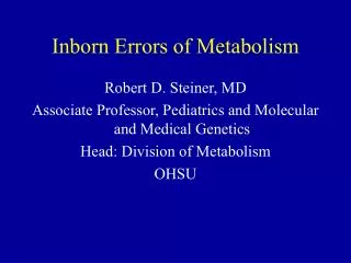
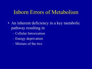
Inborn Errors of Metabolism(IEM) Lecture 2. SDK December 24, 2013. Objectives. Define Inborn error of metabolism Identify the most common errors Explains the mechanism of Inborn error of metabolism. Explain the dietary plan for IEM. SCREEN ABLE IEM . Organic acidemia

E N D
Inborn Errors of Metabolism(IEM)Lecture 2 SDK December 24, 2013
Objectives • Define Inborn error of metabolism • Identify the most common errors • Explains the mechanism of Inborn error of metabolism. • Explain the dietary plan for IEM SDK 2012
SCREEN ABLE IEM • Organic acidemia • Isovalericacidemia • HMG-CoAlyase deficiency • PropionicAcidemia • Methylmalonicacidemia and other • Urea cycle defects • Argininosuccinicaciduria and others • Amino acid disorders • Maple syrup urine disease • PKU • Homocystinuria • Carbohydrate disorders • Galactosemia SDK 2012
4. Amino Acid Disorders SDK 2012
4.1. Phenylketonuria • Phenylketonuria (commonly known as PKU) is an inherited disorder that increases the levels of a substance called phenylalanine in the blood • AutosomalRecessive Disorder. • Inherited error of metabolism caused by deficiency in the enzyme phenylalanine hydroxylase (PAH). • Mutation in both alleles of the gene for the enzyme. • Chromosome 12. • Recessive allele carried by 1 out of every 60 individuals.
Metabolism of PA SDK 2012
Genes related to Phenylketonuria • Mutations in the PAH gene cause phenylketonuria. • The PAH gene provides instructions for making enzyme phenylalanine hydroxylase. • The PAH gene is located on the long (q) arm of chromosome 12 between positions 22 and 24.2. SDK 2012
Metabolism of PA SDK 2012
Clinical features of PKU • Hyperactivity, athetosis, vomiting. • Blond. • Seborric dermatitis or eczema skin. • Hypertonia. • Seizures. • Severe mental retardation. • Unpleasant odor of phenyl acetic acid
Diagnosis &Treatment of PKU • Screening : Guthrie Test. • High Phenylalanine > 20 mg/dl. • High Phenyl pyruvic acid. • DIET. • BH4 (Tetrahydrobioptein).
4.2. Alkaptonuria • Alkaptonuria is an inherited condition that causes urine to turn black when exposed to air. • Ochronosis, a buildup of dark pigment in connective tissues such as cartilage and skin, is also characteristic of the disorder. • This blue-black pigmentation usually appears after age 30. • People with alkaptonuria typically develop arthritis, particularly in the spine and large joints, beginning in early adulthood. • Other features of this condition can include heart problems and kidney stones. SDK 2012
Genes related to Alkaptonuria • Mutations in the HGD gene cause alkaptonuria. • The HGD gene provides instructions for making an enzyme called homogentisateoxidase. • The HGD gene is located on the long (q) arm of chromosome 3 at position 13.33. • This enzyme helps break down the amino acids phenylalanine and tyrosine. SDK 2012
4.2. Alkaptonuria SDK 2012
4.3. Albinism • Deficiency or absence of pigment in the eyes, skin, or hair. SDK 2012
4.3. Albinism SDK 2012
4.4 Homocystinuria • Homocysteine is an intermediate in the metabolism of methionine to cysteine but can also be used to reform methionine. • Homocystineis formed by joining 2 homocysteines • Homocysteine is linked to both folate and vitamin B12 metabolism • Excessive accumulation of homocystine leads to homocystinuria and is caused by • decreased metabolism of homocysteine through either its link to folate Metabolism or through its link to cysteine formation SDK 2012
Genes related to homocystinuria • Mutations in the CBS, MTHFR, MTR, MTRR, and MMADHC genes cause homocystinuria./ • Where is the CBS gene located • The CBS gene is located on the long (q) arm of chromosome 21 at position 22.3. SDK 2012
Methionine Metabolic CycleGene/Enzyme of Transporter MAT I/III Methionineadenosyltransferase I/III GNMT N-Glycinemethyltransferase AHH Adenosylhomcysteinehydrolase CBS Cystathionine B-synthase MTHFR Methylenetetrahydrofolatereductase MMACHC “Methylmalonicaciduria – homocystinuria” C (cblC MMADHC “Methylmalonicaciduria – homocystinuric” D (cblD) LMBRDI cbl F transporter MSRR Methioninesynthasereductase (cblE) MSR Methioninesynthase (cblG)
Homocystinuria (CBS def) • Mental retardation • Ectopialentis • Skeletal abnormalities • Thromboembolism
Diagnosis & Treatment: High methionine and homocystinein blood • TREATMENT: • High dose of B6 and Folic Acid. • Low methionine and high cystine diet, • Betain (trimethylglycine)
5.1. Types of Metabolic Storage Disorders • Glycogen storage diseases (GSD) • Mucopolysaccharidosis (MPS) • Lysosomal storage diseases or lipidosis (LSD) • Peroxisomal diseases
5.2. Glycogen storage diseases (GSD) • Hepatic/ muscle involvement (GSD-III) • Isolated Hepatic involvement (GSD-I, IV & VIII) • Isolated muscle involvement (GSD-V & VII) • Multiple tissues (GSD-II & IV)
Glycogen Metabolism Uridine-Diphosphoglucose 1 Glycogen Straight chains 1 Glycogen synthetase 2 Brancher enzyme (GSD-IV) 2 3 Debrancher enzyme (GSD-III) Glycogen Branched structure 4 Glucose-6-phosphatase (GSD-1) Limit dextrin+ Glucose-1-PO4 Glucose-1-PO4 3 4 Glycogen ( normal branch) + Glucose
Glucosidase/Acid Maltase GSD-II (Pompe Disease) Glycogen D- glucose 1-phosphate
5.3. Galactosemia • Galactosemia is an inherited an autosomal- recessive disorder • deficiency in enzyme (galactose-1-phosphate uridyl transferase) that metabolize galactose • Galactosemia = high level of plasma galactose. SDK 2012
Blood tests Enzyme activity in RBCs Normal range for Galactose-1-phosphate uridyltransferase activity is18.5 to 28.5 U/g Hb. Low blood sugar (hypoglycemia) Urine analysis Reducing substances accumulation (i.e. galactose & galactose-1-P) Diagnosis
Treatment • No pharmacological treatment is currently available • Sources of galactose (especially lactose) must be eliminated from the diet • All dairy products (chesses, yoghurt, ice cream), breast milk, infant formulas, sweeteners • Foods with > 10mg galactose/100g fresh weight must be avoided; dates, papaya, tomatoes, watermelon • Calcium and vitamin supplementation (vitamin D)
6. Mucopolysaccharidosis (MPS) • Are inheritable storage diseases caused by a deficiency of lysosomal enzymes that degrade glycosaminoglycans (GAGs, previously called mucopolysaccharides such as dermatan sulfate, heparan sulfate and keratan sulfate . The MPSs are characterized by the • Intra lysosomal accumulation of GAGs, • Excessive urinary excretion of GAGs, • And variable degrees of progressive mental and physical deterioration • And, in severe forms, premature death. SDK 2012
Mucopolysaccharidosis (MPS) • Types • Seven types Depending on the enzyme deficiency, the metabolism of • Dermatan sulfate, • Heparan sulfate, or • Keratan sulfate may be blocked alone or in combination. • Lysosomal accumulation of the GAGs eventually results in cell, vascular, tissue, and organ dysfunction. SDK 2012
Symptoms & signs Developmental delay. Behavioral dysfunction Coarse facial features & other somatic features Cloudy cornea Abdominal distension (Hepatosplenomegaly) Dysostosis multiplex (Scoliosis and gibbous deformity) Diagnosis Urine for MPS ( heparan , keratan , dermatan) enzyme assay Symptoms & signs (MPS)
Deficiency of iduronidase Accumulation of Dermatan sulfate and heparan sulfate Autosomal Recessive Clinical signs Developmental delay Coarse facial features & other somatic features(large tongue, prominent forehead, Cloudy cornea Hepatosplenomegaly joint stiffness, Hearing loss Hydrocephalus Kyphosis Diagnosis α-Iduronidase deficiency death before 10 yr of age Type 1-Hurler syndrome(MPS-I)
Type II-Hunters • Deficiency of Iduronatesulfatase • The severe form of Hunter syndrome has features similar to those of Hurler syndrome except for the lack of corneal clouding and the slower progression of somatic and central nervous system (CNS) deterioration • X-Linked recessive • Patients with the severe form of MPS II have major deletions or rearrangements of the IDS gene present o Xq28. • Accumulation of Dermatan sulfate and heparan sulfate. • onset of disease usually between 2–4 yr of age • Death usually occurs between 10–15 yr of age
Type IV- Morquio • Deficiency of Galactose-6-sulfatase. • Gene is on chromosome 16q24.3 • Autosomal recessive • Accumulation of Keratan sulfate Characterized • By significant, short-trunk dwarfism, • Fine corneal deposits, • A skeletal dysplasia that is distinct from other mucopolysaccharidoses, • And preservation of intelligence.
7. Lipid Disorder SDK 2012
7.2.Tay-Sachs Disease • Tay-Sachs Disease is rare autosomal recessive genetic disorder . • Genetic mutation on chromosome 15 • Lipid storage disorder that results from deficiency in -hexosaminidase A • Different names are: • GM2gangliosidosis • Hexosaminidase A deficiency • Sphingolipidosis
Symptoms • Loss of hearing • Physical and mental retardation • Seizures • Dementia • And most noticeably detected by the red dots it causes on the retina of an individuals eye
Treatments • Enzyme replacement therapy • Replace with synthetic enzyme • Gene therapy • Replace defective genes • Substrate reduction therapy • Bypass the defect so GM2 can be metabolized
8. Peroxisomal Disorder • Peroxisomal disorders are a group of genetically heterogeneous metabolic diseases that share dysfunction of peroxisomes. • Peroxisomes are cellular organelles that are an integral part of the metabolic pathway. • They participate in important peroxisome-specific metabolic pathways, such as beta-oxidation of very-long-chain fatty acids (VLCFA) and detoxification of hydrogen peroxide. • Peroxisomes are also involved in the production of cholesterol, bile acids,platelet activating factor [PAF] and plasmalogens, which contribute to a big part of the phospholipid content of the brain white matter.
Peroxisomal Diseases Adrenoleukodystrophy: Deficiency in -oxidation of very long- chain fatty acids Zellweger syndrome: Defect in protein import, giving rise to “ghost peroxisomes”
8.1.Zellweger syndrome • Cerebro-hepato-renal syndrome of Zellweger (Zellweger syndrome) is a peroxisomal disease that is biochemically characterized by abnormal accumulation of very long chain fatty acid. • most severe form of peroxisomal disorder due to errors in peroxisomal biogenesis or defects in maintaining peroxisomalintergrity. SDK 2012