Download
1 / 57
570 likes | 898 Vues
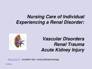
Nursing Care of Individual Experiencing a Renal Disorder: Vascular Disorders Renal Trauma Acute Kidney Injury. Renal A & P -excellent site- renal pathophysiology. I. A&P of the Kidney- (locate structures). Fibrous capsule Renal cortex Renal medulla Pyramids Papillae Minor calyx

E N D
Nursing Care of Individual Experiencing a Renal Disorder:Vascular DisordersRenal TraumaAcute Kidney Injury Renal A & P -excellent site- renal pathophysiology
I. A&P of the Kidney- (locate structures) • Fibrous capsule • Renal cortex • Renal medulla • Pyramids • Papillae • Minor calyx • Major calyx • Renal pelvis • Ureter
II. Functions of the Kidneys • Regulates ______ & _________ of extracellular fluid • Regulates fluid & electrolyte balance thru processes of: glomerular__________, tubular _________, and tubular _____________. Name some of the F & Es regulated by kidneys __________________ volume and composition filtration, reabsorption, secretion Na, K, HCO3, H,
Functions of the Kidneys (cont) • Regulates acid-base balance through • *Hormonal functions: (BP control), multisytem effect through > • HCO3 and H+ • Renin Release RAAS= Renin-Angiotensin-Aldosterone-System
How the RAAS Pathway Works (Know this) Click here- Renin-Angiotensin-Aldosterone Pathway The Song! Valerie Kolmer 2006
Quick Quiz Pick the correct pathway of the RAAS • 1. Renin – Angiotensin II – ACE – ADH – Aldosterone • 2. Renin – Angiotensin I – Aldosterone – ADH –ACE • 3. Renin-Angiotensin 1-ACE-Angiotensin 11-Aldosterone #3
Functions of the Kidneys (cont) • Erythropoietin Release • If a patient has chronic renal failure, what condition will occur/WHY??? *Anemia – from impaired erythropoietin production and platelet abnormalities > bleeding risk
Functions of the Kidneys (cont) • Activated Vitamin D • Necessary to absorb Calcium in the GI tract. If a patient has renal failure, what will happen to the patient’s serum calcium level? __________________ *Low serum calcium level/elevated phosphate- inability kidneys to activate vit D- *hypocalcemia > parathyroid gland > secretes PTH > stimulates bone demineralization > release calcium from bones!
Review: Functions of the Kidneys • Regulate • 1.___________ • 2.___________ • 3.___________ • 4.___________ • Release of ________________ • Activation of _______________ 1. volume 2. composition extrcellular fluid 3. fluid 4. electrolyte balance 1. renin 2. activation Vit D
III. Nephron- functional unit of Kidney! • How the Nephron Works! Click-watch YouTube video!
Identify the Nephron’s Parts • Glomerulus • Bowman’s capsule • Proximal tubule • Loop of Henle • Distal tubule • Collecting duct Click here for Nephron A&P & Games too!
Renal Trauma(click- view renal trauma slides) ` • Etiology: • Blunt force from falls, MVA, sports injuries, knife/gunshot wounds, impalement, rib fractures • Common Manifestations: • Microscopic to gross hematuria • Flank or abdominal pain • Oliguria or anuria • Localized swelling, tenderness, ecchymosis flank • area (Turner’ssign=bluish discoloration flank area • due to retroperitoneal bleeding) • ( Cullen's sign -periumbilical ecchymosis; figure A ; Grey-Turner's sign -ecchymosis • on the abdominal flank; figure B ) and ascites. ) • Sign/Symptoms depend upon severity injury • *Severe blood loss/signs shock
Renal Trauma Major injury to the renal hilar vessels with devascularization of the kidney is a grade V injury
Renal Trauma • What are common diagnostic tests used in renal trauma? • *UA (hematuria)- • H & H (decreasing values) • *CT, MRI, IVP with cystography • Ultrasound; maybe renal arteriogram CT-determine if peritoneal violation and predict need for laparotomy-here initially see extravasation and fluid in paracolic gutters (peritoneal violation) and also a hematoma in perirenal space
Renal Trauma-Interventions • Minor or Major? • Conservative • Bedrest and close observation. • Monitor for S & S of ? Bleeding, potential Shock!! (Hypovolemic) Monitor H & H
Renal Trauma-Interventions • Surgical- • Surgical repair, maybe nephrectomy • Percutaneous arterial embolization during angiography • Nursing management • Accurate assessment • Monitor H & H levels • Bedrest; close observation; evaluate S & S of shock • Fluid mgt • Prevent complications/monitor I & O • Manage drainage tubes • **Daily weights
Renal Surgery-Nephrectomy • Indications for Nephrectomy: • Renal tumor • Massive Trauma • Polycystic Kidney Disease • Donating a Healthy kidney
Renal Surgery-Nephrectomy • Post Op Nursing Management • Strict I & O • Urine output should be at least _____. • What should u.o. be if patient had bilateral nephrectomy? ______. • Observe urine • TCDB & incentive spirometry • Incision in flank area, 12th rib removed • Medicate for pain as ordered 30 ml/hr I hope you said “0”
Renal Vascular ProblemsPatho of HTN-Nephrosclerosis • Development of arterio sclerotic lesions in the arterioles and glomerular capillaries ↓ Decreased blood flow which leads to ischemia and patchy necrosis ↓ Destruction of glomeruli ↓ Decrease in GFR
Vascular Disorders of the KidneyRenal Artery Stenosis • Definition: Narrowing of one or both renal arteries due to atherosclerosis or structural abnormalities. • Common Manifestation! • Uncontrollable HTN- Why? • Think “pathophysiology” of HTN!
Vascular Disorders of the KidneyRenal Artery Stenosis • Treatment/Collaborative Care • Diagnostic Tests • Renal arteriogram-most definitive • Management • Conservative-antihypertensive meds • Percutaneous Transluminal Angioplasy • Surgical re-vacularization (Graft) • ?Nephrectomy
Vascular Disorders of the KidneyRenal Artery Stenosis • Treatment/Collaborative Care What type of procedure is this? What are some post procedure nursing care interventions?
Vascular Disorders of the Kidney • Renal Vein Stenosis • Definition: Partial occlusion in one or both renal arteries due to atherosclerosis or structural abnormalities in vein by a thrombus. • Risk Factors: • Nephrotic syndrome • Use of Birth control pills • Certain Malignancies
Vascular Disorders of the KidneyRenal Vein Thrombosis/Occlusion • Pathophysiology/etiology • Cause unclear-thrombus forms in renal vein • Associated with trauma, nephrotic syndrome gradual • deterioration of renal function • Common Manifestations/Complications • Decreased GFR • Signs of Renal Failure • **Complication ---*Pulmonary Embolus
Vascular Disorders of the KidneyRenal Vein Thrombus/Occlusion • Treatment/Collaborative Care Diagnosis- Renal venography Management Thrombolytic drugs- streptokinase or tPA Anticoagulant therapy to prevent further clot formation
Acute Kidney Injury • Definition: • Rapid decline in renal function- leads to accumulation of nitrogenous wastes (azotemia) • Kidneys unable to remove urea from blood-become uremic (multiple body symptoms affected) Click here-MD lecture on AKI YouTube 8 min!
Acute Kidney Injury Etiology of AKI: • Pre-renal • Intra-renal • Post renal
Causes of “pre-renal” AKI failure-all related to dec. blood flow to kidneys! Hypovolemia: dehydration, shock, burns Decreased cardiac output: CHF, MI, arrythmias Dec. vascular resistance (septic shock, etc) Renal vascular obstruction: renal artery stenosis, thrombus. Etiology of Acute Kidney InjuryPre-renal (most common cause AKI!)
Direct injury to the kidneys/nephrons Due to renal tissue causing damage to renal tissue (parenchyma) Causes of “intra renal” AKI failure: Acute ischemia (prolonged pre-renal) ATN (acute tubular necrosis) *Destruction of tubular epithelial cells, slough, plug tubules- abrupt decline in renal function-recovery possible if basement membrane remains intact & tubular epithelium regenerates) Etiology of Acute Kidney InjuryIntra-renal
Hemolytic blood transfusion (ATN) Trauma (crush injuries > release myoglobin; damage muscle tissue > blocks tubules (rhabdomylosis)*know (ATN) Nephrotoxic drugs/chemicals (ATN) Aminoglycosides* Radiographic contrast agents Arsenic, lead, carbon tetachloride Acute glomerulonephritis/pyelonephritis Systemic lupus Causes of Intrarenal Failure
Renal ischemia Disruption basement membrane;destruction tubular epithelium Nephrotoxic agents Necrosis tubular epithelium… plug tubules; basement membrane intact. Potentially reversible IF Basement not destroyed and tubular epithelium regenerates Causes of Acute Kidney Injury (ATN) Renal ischemia Nephrotoxic agents
Etiology of Acute Kidney Injury Post-renal • Causes of “post-renal failure” (mechanical obstruction of urinary outflow; urine backs up into renal pelvis) • BPH (Benign Prostatic Hypertrophy) • Calculi • Trauma • Prostate cancer
BUN (blood urea nitrogen) Normal = 8-20 mg/dl; measurement of amt of urea in blood What is urea? BUN fluctuates BUN elevated in? BUN increased when? Diagnostic Tests in Acute Kidney Injury: Nitrogenous waste product from breakdown of (protein) amino acids by liver - Excessive protein intake, GI bleeding, dehydration, Overhydration, primary liver disease (synthesis of urea depends upon liver), kidney disease
Question? • Which urinary symptoms is the most common initial manifestations of AKI? a- dysuria b- anuria c- hematuria d- oliguria Oliguria-dec. GFR, less than 400 ml/24 hr
Question… • The client’s BUN is elevated in AKI. What is the likely cause of this finding? • a- fluid retention • b- hemolysis of red blood cells • c- below normal protein intake • d- reduced renal blood flow Elevated BUN reflects impairment of renal function, thus indicates reduced renal blood flow- normal blood flow is needed for adequate GFR. Also-Hemolysis of Red blood cells will obstruct renal tubules can also cause an elevated BUN
Serum Creatinine:end product of muscle and protein metabolism; excreted by the kidneys at a constant rate Normal = 0.6-1.2 mg/dl Directly related to GFR 2 X normal (2.4) = 50% nephron fx loss 10 X normal (12) = 90% nephron fx loss MORE ACCURATE INDICATOR of RENAL FUNCTION THAN BUN BUN; Creatinine ratio Normal= 10:1 BUN Creatinine 16 1.6 12 1.2 8 0.8 Diagnostic Tests in Acute Kidney Injury:
Creatinine clearance Most accurate indicator of Renal Function Reflects GFR Involves a 24 hr urine/serum creatinine Formula: Amount of urine creatinine X urine V serum creatinine Normal= 100-135ml/minute Diagnostic Tests in Acute Kidney Injury:
Urine Specific Gravity Normal= 1.003-1.030 Fixed sp. Gravity- 1.010 usually in AKI kidneys lose ability to concentrate urine Serum Electrolytes 1- Serum Sodium Normal= 135-145meq/L May be high, low, or normal High in Volume deficit (dehydration) Low due to damaged tubules not conserving sodium Diagnostic Tests in Acute Kidney Injury:
Serum Electrolytes 2- Serum K+ Normal= 3.5-5.5 meq/l Almost always increased WHY? Kidneys excrete 80-90% of our K+ If K+> 6.0; treatment initiated to prevent? Diagnostic Tests in Acute Kidney Injury: > inability kidneys to excrete potassium; *acidosis worsens hyperkalmeia –hydrogen ions enter cells; potassium driven out of cells!! *Fatal dysrhythmias!
Serum Electrolytes 3- Serum Calcium Normal= 9-11mg/dl due to production of activated Vitamin D; needed to absorb calcium from GI tract What other process is occurring to decrease serum calcium??? Diagnostic Tests in Acute Kidney Injury: *Hypocalcemia causes PTH to be released > bone demineralization > more phosphate be released from bone; *phosphate already high, calcium binds with phosphates
Serum Electrolytes 4- Serum Phosphorus Normal= 2.0-4.5mg/dl Phosphorus- product of protein breakdown excreted by the kidneys What other process is occurring to increase serum phosphorus? Diagnostic Tests in Acute Kidney Injury: Parathyroid gland secretes PTH because calcium is low, causes phosphorus also to be released from bone > phosphorus
ABGs pH Metabolic acidosis due to ability of kidneys to excrete acid metabolites (uric acid, ammonia) so the pH will be __________. Also, bicarb levels due to bicarb being used up to buffer excess H+ ions. Diagnostic Tests in Acute Kidney Injury: low
Stages of Acute Kidney Injury • Oliguric Phase • Usually appears 1-7 days of initiating event • Diuretic Phase • Start varies, usually within10-12 days of onset oliguric phase • Recovery • Usually within a month, recovery takes up to 12 months
Oliguric Phase Onset: 1-7 days Duration: 10-14 days Urine output: Less than 400 ml/24 hours in 50% of patients Signs and Symptoms to Anticipate? (similar to CKD) Urine less that 400 ml in 24 hours Specific gravity fixed at 1.010 in oliguria in intra renal failure Fluid overload Urine with RBCs, casts, WBCs K likely elevated Oliguric Phase of AKI:
Acute Kidney Injury: Oliguric • Metabolic acidosis: • kidneys unable to synthesize NH3, need H excretion, or excrete acid metabolites; serum bicarbonate, dec. used to buffer H (Kussmaul breathing) • *Ca deficit; phosphate excess; • dec. GI absorption Ca (lack of active vitamin D) *not significant in AKI • Nitrogenous product accumulation: • unable to eliminate urea and creatinine > elevated BUN, serum creatinine
Onset: days to weeks Duration: 10 days (1-3 weeks) Urine output:1-3 liters/day Signs and Symptoms to Anticipate? Fluid Volume Overload or Fluid Volume Deficit Elevated BUN and serum creatinine (continue) K likely to be elevated or decreased??? Hyponatremia and hypotension Diuretic Phase of AKI: Deficit! Decreased
Recovery Phase Onset: When BUN and Creatinine are stablized Duration: 4-12 months Urine output: Normal Signs and Symptoms to Anticipate? Continue to monitor for signs and symptoms of F & E imbalances All body systems for effects of fluid volume changes Recovery Phase of AKI:
Initial Treatment-Oliguric Phase • Fluid Challenge/Diuretics • Done to r/o dehydration as cause of AKI and “ blast out tubules” if ATN. • 250-500cc NS given I.V. over 15 minutes • Mannitol (osmotic diuretic) 25gm I.V. given • Lasix 80mg I.V. given • Should see what within 1-2 hours???? Urine- at least 30ml/hr!)
Treatment During: OliguricPhase • If fluid challenge fails, fluid intake is usually limited and client is placed on fluid restriction • Restriction is limited to 600ml + u.o. past 24 hours • Physician will specify in the orders how much. Question: Patient’s u.o. on Tuesday=300ml, what will be his fluid intake allowed on Wednesday? ________ 900 ml (600 + 300)
Acute Kidney Injury: Management of…. • 1- Treat primary disease/condition whether it is pre-intra-or post renal problem. • 2-Prevention: • Frequent monitoring for early signs of AKI in at risk patients • What can the nurse assess for at this point? • 3-Assess for Fluid V deficit vs Fluid V overload • Strict I & O • Daily weights 500ml-=1 lb. (1kg = approx 1000ml fluid) • Monitor lab values…which ones? _______