Prolapse Repair
Prolapse Repair. Extracted from: Turner and McIlwraith's Techniques in Large Animal Surgery, 4th Edition - Dean A. Hendrickson and A. N. ( Nickie ) Baird. Retention Suturing of the Bovine Vulva ( Buhner’s Method). Relevant Anatomy – refer to Cmap. Indications

Prolapse Repair
E N D
Presentation Transcript
Prolapse Repair Extracted from: Turner and McIlwraith's Techniques in Large Animal Surgery, 4th Edition - Dean A. Hendrickson and A. N. (Nickie) Baird
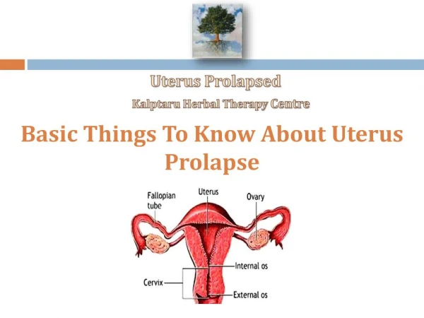
Retention Suturing of the Bovine Vulva (Buhner’s Method) Relevant Anatomy– refer to Cmap Indications Vaginal or cervical prolapse occurs with the greatest frequency during the last trimester of gestation in cows. The condition may also occur during early postpartum or estrus, however. Prolapses are usually classified by the duration of the condition and the extent of the prolapse. For example, first-degree vaginal prolapse involves only intermittent exposure of the vaginal floor, usually occurring when the cow is lying down. Second degree vaginal prolapse infers that the vaginal floor is continuously exposed. The urinary bladder may or may not be retroflexed to be included in the prolapsed tissue but urination may be impeded if the urethra becomes occluded.
Indications Cont’d Third degree vaginal prolapses involve a continuous exposure of the vaginal floor and the cervix through the vulva. Third degree prolapses in Bos indicus and Bos taurus breeds have been differentiated based on the observation that third degree prolapses in Bos indicus are usually primary prolapses of the cervix that have not progressed from a first- or second-degree vaginal prolapse. The Bos taurus breeds, however, usually will progress from a first- or second-degree prolapse to a third-degree prolapse. Of the prolapsed tissue, the cervical os is usually located most dorsally and an extremely edematous vaginal floor most ventrally. A fourth-degree prolapse is described as either a first- or second-degree prolapse of a long enough duration that the prolapsed tissue has become necrotic.
Indications Cont’d The buried purse-string suture (Bühner method) is a simple and effective way to retain vaginal or uterine prolapse in the cow. The method consists of a deeply placed circumferential suture that effectively simulates the action of the constrictor vestibular muscle. The purse-string suture may be permanent or temporary. It is strong and does not tear out as frequently as externally placed suture patterns (lacing, Halsted, and quill). The Minchev technique is used to anchor the anterior dorsal vagina to the gluteal area by passing heavy suture through the anterior dorsal vaginal wall, the sacrosciatic ligament, and the skin in the gluteal area. This technique does not restrict the vaginal opening like the Bühner method but may still permit prolapse of the vaginal floor or result in necrosis of the dorsal wall in which the sutures are passed.
Anesthesia and Surgical Preparation The cow should be restrained in a chute or crush, and some cows may be recumbent during the procedure. The surgery is performed with the animal under caudal epidural analgesia. Following administration and onset of the epidural analgesic, the perianal area and prolapsed tissues are cleaned and treated with an antiseptic. Osmotic agents and massage may then be used to reduce the size of the prolapse. Some cases with significant edema in the prolapsed tissue will benefit from a short-term pressure wrap and dorsal support to help alleviate the edema. If the bladder is included in the prolapsed vagina such that the urethra is obstructed, one should lift the prolapsed vagina dorsally which will straighten the urethra and allow the bladder to empty thus decreasing the size of the prolapse. Instrumentation General surgery pack Bühneror Gerlachperivaginal needle Perivaginalsuture tape or sterile, 1-cm (half-inch) umbilical tape
Surgical Technique A typical prolapse is depicted in Figure 14.10A. The prolapse is reduced, the vagina is returned to its correct anatomic location, and the perianal area is scrubbed once again. A transverse skin incision about 1 cm long is made midway between the dorsal commissure of the vulva and the anus. Another horizontal incision is made about 3 cm below the ventral commissure of the vulva. The perivaginal needle is introduced into the ventral skin incision and is driven perivaginally through the deep subcutaneous tissues parallel to the vulva. One hand is placed in the vagina to guide the needle. The needle should be driven as deep as possible (about 5–8 cm) and directed out the dorsal skin incision (Figure 14.10B).
Surgical Technique Cont’d A piece of sterile perivaginal suture tape (or sterile umbilical tape) soaked in a suitable antibiotic solution, is threaded through the eye of the needle and is drawn down to emerge through the ventral skin incision (Figure 14.10C). At the same time, the tape is held at the dorsal incision, so the end is not lost in the tissue. The tape is then removed from the needle, and the needle is threaded up the contralateral side of the vulva (about 5–8 cm) to emerge through the dorsal incision. The tape is threaded into the eye of the needle once again (Figure 14.10C), and then the needle is withdrawn ventrally, resulting in two free ends of tape emerging from the ventral skin incision.
Surgical Technique Cont’d The two free ends of the tape are tied, ensuring that the loop of tape at the dorsal incision is buried (Figure 14.10D). The tape is tied so the resulting suture encircling the vulva will admit 2–3 fingers. If a square knot is used to anchor the tape, the knot will bury itself. This minimizes the chances of contamination of the suture material and thereby avoids a wicking effect with the suture and secondary infection. The dorsal and ventral incisions may be closed with a simple interrupted suture of nonabsorbable material, to further decrease the chances of secondary infection around the umbilical tape. If the cow is close to calving, we recommend that the tape be secured in the ventral incision with a bow knot. This knot allows the suture to be removed or, at least, undone to reduce tension at the time of parturition. One of the other methods to retain the prolapse may also be used. One should also consider two separate stab incisions ventrally for placement of the needle if the cow is pregnant so the suture is not buried and can be easily untied or cut. In pregnant cows that will be returned to the range or pasture to calve, we use a double strand of gut suture instead of Bühner tape. The gut suture will break at parturition while the Bühner tape will not. The disadvantage of using the gut suture for this situation is that the suture may fail prior to calving.
Postoperative Management The cow requires close observation to time removal or loosening of the suture correctly in relationship to parturition. The knot should be untied, and the vulva should be gently dilated to reduce tension on the suture. Complications and Prognosis The Bühner method of repair for vaginal prolapse offers secure retention of the vagina and cervix with the convenience of quick release during calving. Calving through Bühner sutures is one of the most severe complications of this procedure and can result in severe lacerations and damage to the vulva and perineal area. Certain cattle that have a pendulous vulva may be predisposed to edema, swelling, and even necrosis of the vulva following Buhner method of repair due to the increased tension required to retain the vagina and cervix.