AJCC Staging Moments
221 likes | 404 Vues
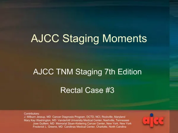
AJCC Staging Moments. AJCC TNM Staging 7th Edition Rectal Case #3. Contributors: J. Milburn Jessup, MD Cancer Diagnosis Program, DCTD, NCI, Rockville, Maryland Mary Kay Washington, MD Vanderbilt University Medical Center, Nashville, Tennessee

AJCC Staging Moments
E N D
Presentation Transcript
AJCC Staging Moments AJCC TNM Staging 7th Edition Rectal Case #3 • Contributors: • J. Milburn Jessup, MD Cancer Diagnosis Program, DCTD, NCI, Rockville, Maryland • Mary Kay Washington, MD Vanderbilt University Medical Center, Nashville, Tennessee • Jose Guillem, MD Memorial Sloan-Kettering Cancer Center, New York, New York • Frederick L. Greene, MD Carolinas Medical Center, Charlotte, North Carolina
Rectal Case # 3 Presentation of New Case • Newly diagnosed rectal cancer patient • Presentation at Cancer Conference for treatment recommendations and clinical staging
Rectal Case # 3 History & Physical • 51 yr old male who presented with diarrhea and abdominal pain • No family history of colon or rectal cancer
Rectal Case # 3 Imaging & Endoscopy Results • Colonoscopy-single large circumferential fungating mass in rectum 2 to 5cm from anus Retroflexed endoscope with rectal tumor used with permission Julio Murra-Saca, MD El Savador Atlas of Gastrointestinal Video Endoscopy
Rectal Case # 3 Diagnostic Procedure • Procedure • Biopsy rectal mass • Pathology Report • Adenocarcinoma, invasive • Grade 2 • CEA: 4.3
Rectal Case # 3 Imaging Results • Endorectal ultrasound • Invasion into peri-rectal fat • CT chest/abd/pelvis • Thickening in rectal and perirectal soft tissue • 1.2cm peri-iliac node mesenteric node • 1.7cm perirectal lymphadenopathy • No liver metastases used with permission Julio Murra-Saca, MD El Savador Atlas of Gastrointestinal Video Endoscopy
Rectal Case # 3 Clinical Staging • Clinical staging • Uses information from the physical exam, imaging, and diagnostic biopsy • Purpose • Select appropriate treatment • Estimate prognosis
Rectal Case # 3 Clinical Staging • Synopsis- patient with rectal mass and perirectal tissue involvement, and clinically involved nodes • What is the clinical stage? • T____ • N____ • M____ • Stage Group______
Rectal Case # 3 Clinical Staging • Clinical Stage correct answer • T3 • N1 • M0 • Stage Group IIIB • Based on stage, treatment is selected • Review NCCN treatment guidelines for this stage
Rectal Case # 3 Clinical Staging • Rationale for staging choices • T3 for into non-peritonealized pericolic or perirectal tissues • N1 because nodes were clinically positive on imaging • M0 because there was nothing to suggest distant metastases; if there was, appropriate tests would be performed before developing a treatment plan
Prognostic Factors Clinically Significant • Applicable to this case • CEA: 4.3 • There are no prognostic factors required for staging
Rectal Case # 3 Surgery & Findings • Patient received neoadjuvant Rx with chemotherapy and radiation therapy • Procedure • Transabdominal resection • Operative findings • Colo-anal anastomosis performed with 2 cm margin
Rectal Case # 3 Pathology Results • Residual adenocarcinoma, rectum • Tumor size - 3cm • Grade 3, poorly differentiated • Through muscle wall into pericolonic soft tissue • Margins negative • Circumferential resection margin clear by 8mm • Mets 3/12 regional nodes • No perineural or lymph-vascular invasion • Tumor deposits were not identified
Rectal Case # 3 Pathologic Staging • Pathologic staging • Uses information from the clinical staging supplemented or modified by information from surgery and the pathology report • yp is assessment at conclusion of therapy • Purpose • Additional precise data for estimating prognosis • Calculating end results (survival data) • yp – extent of response to therapy
Rectal Case # 3 Pathologic Staging • Synopsis- patient with rectal ca into pericolonic soft tissue, and positive nodes after neoadjuvant chemo and RT • What is the pathologic stage? (remember, clinical M may be used in pathologic staging) • T____ • N____ • M____ • Stage Group______
Rectal Case # 3 Pathologic Staging • Pathologic Stage correct answer • ypT3 • ypN1 • cM0 • ypStage Group IIIB • Based on pathologic stage, there is more information to estimate prognosis and adjuvant treatment is selected
Rectal Case # 3 Pathologic Staging • Rationale for staging choices • ypT3 for into non-peritonealized pericolic or perirectal tissues after neoadjuvant chemo/RT • ypN1 because nodes were positive on exam after neoadjuvant chemo/RT • cM0 - classified by M status prior to therapy • y prefix used to show stage during or following neoadjuvant therapy
Prognostic Factors Clinically Significant • Applicable to this case • CEA: 4.3 • Circumferential resection margin: 8mm • Tumor deposits: no • Perineural invasion: no • There are no prognostic factors required for staging
AJCC Cancer Staging Atlas T3 into non-peritonealized pericolic or perirectal tissues (adventitia)
Rectal Case # 3 Recap of Staging • Summary of correct answers • Clinical stage T3 N1 M0 Stage Group IIIB • Pathologic stage ypT3 ypN1 cM0 ypStage Group IIIB • The staging classifications have a different purpose and therefore can be different. Do not go back and change the clinical staging based on pathologic staging information.
Staging Moments Summary • Review site-specific information if needed • Clinical Staging • Based on information before treatment • Used to select treatment options • y Pathologic Staging • Based on clinical data PLUS surgery and pathology report information after neoadjuvant therapy • Assesses response to treatment • Used to evaluate end-results (survival)