Understanding Tissue-Specific Gene Expression: The Role of Nuclear Matrix
80 likes | 194 Vues
This teacher's guide focuses on the concept of tissue-specific gene expression and the unique functions of cells despite sharing identical DNA. Through a series of five engaging exercises, students explore how different tissues utilize specific genes and the impact of the nuclear matrix on gene accessibility. By encouraging classroom discussions, teachers can help students differentiate between housekeeping genes and tissue-specific genes, examine cell size, and understand DNA organization in the nucleus. The guide promotes active learning and adaptability to different teaching styles.

Understanding Tissue-Specific Gene Expression: The Role of Nuclear Matrix
E N D
Presentation Transcript
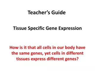
Teacher’s Guide Tissue Specific Gene Expression How is it that all cells in our body have the same genes, yet cells in different tissues express different genes?
Throughout this video students are provided with 5 exercises aimed to demonstrate, towards the conclusion of the video, the role of the nuclear matrix in exposing a gene or hiding it and how that contributes to regulating tissue specific gene expression. The teacher is expected after each exercise to spend time with the students analyzing their answers and discussing with them the outcome of their answers before moving on with the video towards the next exercise. In the upcoming slides, recommendations are made on how best to analyze the outcome of each exercise, but individual teachers may find better ways, and are encouraged to use what they see best fits their students.
Exercise I: Since all cells have the same genes then how do they acquire different functions and even look different? • To answer this, students can break into groups or discuss the possible answers with their teacher. Here the students ought to become aware, if not already aware, of the difference between house keeping genes and tissue specific regulated genes. • The expression of the latter imparts on a cell specific functions and shapes. This is illustrated in the example of the four cells that are shown in the video. Namely, breast cells, inner ear cell, neuron, sperm and ova. • Note: The images for these cells are colored due to fluorescence-based imaging technology (as in breast and neuron) or visualized via scanning electron microscopy (as in inner ear cell, ova and sperm). The latter allows for a greater degree of magnification.
A basic notion in biology that most high school students fail to conceptualize is the fact that all cells in the animal or human body contain the same DNA yet different cells in different tissues express different genes. Genes can be broadly divided into two classes: 1- “House-keeping genes”. These are needed, to sustain basic functions of a cell such as nutrient uptake, protein synthesis, DNA synthesis and other basic metabolic functions. 2- “Tissue Specific Genes”. The expression of these genes vary depending on the type of tissue and the stage of development. These genes are responsible for the synthesis of proteins specifically produced by one cell/tissue type but not by another, and at a certain stage of development. (Milk protein in breast cells, insulin in pancreatic cells etc …)
Exercises II & III: How big is the cell? • In the second exercise, students are asked four questions that basically alerts them to the fact that the cell can not be seen with the naked eye, and that it’s about 20 micrometers in size. • In the third exercise, students will be able to guess the size of all cellular organelles by sketching to scale the cell and benchmarking the size of the organelles to the actual size of the 20 micrometer cell. • The intent of these two exercises is to allow the teacher to guide students into the nucleus while appreciating the small size of this organelle. Nevertheless and despite its size (10 micrometers or so), the nucleus can efficiently package the entire DNA in a manner that is highly organized as will become evident to the students as they proceed through the video. • Note: For exercise II, it may be a good idea to tabulate the student responses and show them how many got the answers right vs. those that got it wrong. This can be followed by discussion of the results. • In Exercise III, the teacher should be after approximate values for the size of organelles rather than specifics. For example a nucleus is about 10 microns, a mitochondria is about 1 micron, the membrane thickness is few nanometers so on and so forth. These measures will alarm the students as they learn that the DNA is 2 meters in length and has to be packaged in an orderly manner inside the nucleus.
Exercise IV: DNA packaging and organization in the nucleus. • In this exercise students will most likely answer the questions posed correctly. i.e. that the DNA packaging is not random and that it’s packaging differs between tissues. • At this point, it is a good idea to discuss with the students how such an organization can be achieved, maintained, and modulated. The teacher can at this point suggest that the existence of a “tiny” scaffold in the nucleus (later to be identified to the students as a nuclear matrix also known as a nuclear skeleton) will allow the cell to achieve the packaging and organization it needs.
Exercise V: How does the cell expose and or hide the genes that it needs to express or shut down? • In this exercise, it is important that students understand that the nuclear matrix helps in the organization and packaging of the DNA. In addition and most importantly, students should be aware that that the nuclear matrix is also a dynamic structure. As it changes its architecture, the DNA also changes its mode of packaging thus exposing or hiding genes of interest. • Note: As a teacher, please remember that other factors also affect DNA packaging and gene expression. Example are histone and non-histone proteins and DNA methylation (epigenetics) but these topics were not discussed in this presentation. This was mentioned in passing at the closing of the video prior to the concluding remarks. • Next slide demonstrates in a schematic diagram what a nuclear matrix looks like. A transmission electron microscopy image of the matrix is also shown.
The Nuclear Matrix Nuclear membrane Cytoskeleton Nuclear Matrix Electron microscopy image of the nuclear matrix network inside the nucleus, bound by the nuclear membrane. The cell cytoskeleton appears in the cytosol. Schematic diagram of a nuclear matrix referred to as inner and outer matrix. From: Gene TherMolBiolVol 13, 231-243, (2009); by Linnemann, and Krawetz