Download
1 / 88
950 likes | 3.2k Vues
Special Senses. Ch 15. The Special Senses. Taste Smell Vision Hearing Balance. Taste. Taste and smell are involved with specific receptor cells called chemoreceptors . respond to chemicals in an aqueous solution. food dissolved in saliva.

E N D
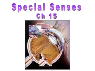
Special Senses Ch 15
The Special Senses Taste Smell Vision Hearing Balance
Taste Taste and smell are involved with specific receptor cells called chemoreceptors respond to chemicals in an aqueous solution food dissolved in saliva airborne chemicals dissolved in mucous membrane
Taste Buds Filiform papilla Fungiform papilla Circumvallate Papilla Tongue epithelium Connective tissue
Five Basic Tastes Why are they important? Salty- metallic ions Umami- savory/meaty Bitter- alkaloid Sour- H+ Sweet- sugar
Experiment Dry tongue with a paper towel and place a little sugar on surface. What do you taste?
Gustatory pathway Facial nerve (afferent) 2/3 anterior portion of tongue Glossophyngeal posterior 1/3 of tongue Vagus nerve- few taste buds on epiglottis an pharynx These afferent fibers synapse in medullathalamus gustatory cortex in parietal lobes and fibers to hypothalamus in limbic system
Tastetriggers reflex involved in digestion; causes an increase of saliva in mouth (amylase) and gastric juice in stomach acids cause strong salivary reflex bad tasting food causes gagging or reflexive vomiting taste can change over time taste is 80% smell Mouth also contains: Thermoreceptors Mechanoreceptors Nociceptors- sensitive nerve fibers that are aware of painful stimuli
Olfaction Smell not as good as animals; however, some people are wine tasters, perfumers If you smell a particular odor all day, you won’t recognize its presence, you become accustomed, ex. garbage men Old people lose sense of smell- lots of perfume Humans can distinguish 10,000 or so chemicals What we really smell is pain: ex. chili, ammonia, menthol (cold) Specific chemicals cause specific patterns of neurons to fire
Olfaction Olfactory epithelium Olfactory tract Olfactory bulb Nasal conchae Route of inhaled air (a) Figure 15.21a
Olfaction Mitral cell (output cell) Olfactory tract Glomeruli Olfactory bulb Cribriform plate of ethmoid bone Filaments of olfactory nerve Lamina propria connective tissue Olfactory gland Axon Basal cell Olfactory receptor cell Olfactory epithelium Supporting cell Dendrite Olfactory cilia Mucus Route of inhaled air containing odor molecules (b) Figure 15.21a
Anosmias loss of sense of smell Lose sense of smelllose taste May be genetic or a cold (mucus), allergy, zinc deficiency Uncinate- olfactory hallucinations; may be psychological ex. rotting meat smell Olfactory auras- prior to epileptic attack
sclera tear drainage canal iris pupil The Eye palpabre cornea Lacrimal caruncle lateral commisure Medial commisure palpabre
Pupil bright light normal light dim light
Lacrimal Apparatus FLOW OF TEARS Lacrimal gland Lacrimal ducts Sup. or inf. lacrimal canal Lacrimal sac Nasolacrimal duct Nasal cavity
Extrinsic Eye Muscles Superior oblique Superior rectus opticnerve Medial rectus Inferior oblique Lateral rectus Inferior rectus
Fibrous Tunic • Fibrous tunic- sclera and cornea (outer most layer) • Cornea • 100s of sheets of collagen fibers between sheets of epithelium and endothelium • Clear because regular alignment • Role in light bending • Avascular but does have pain receptors • Regenerates
Vascular Tunic • Vascular tunic- uvea: choroid, cilliary body, iris, pupil (middle layer) • Choroid- rich vascular nutritive layer; contains a dark pigment • that prevents light scattering within the eye • Cilliary body- lens is attached; contains muscles that change the • lenses shape • Iris- pigmented ring of muscular tissue composed of circular • and radial muscles • reflex contraction of circular muscle in bright light (small dia of pupil) • reflex contraction of radial muscle in dim light (large dia of pupil) • Pupil- central hole in iris
Sensory Tunic • Sensory tunic- retina (inner most layer) • Photoreceptors: • rods (dim light, contains pigment rhodopsin) 120 million rods • Cones (color vision, not evenly distributed, concentrated in fovea) 6 million cones • Optic disc- blind spot because its where optic nerve leaves the eyeball (no rods or cones) • Macula lutea- yellow spot, area of high cone • Fovea centralis- in center of macula lutea, contains only cones, area of greatest visual acuity
Vitreous Humor • Vitreous humor- behind lens, gel-like substance with fine collagenic fibrils imbedded in as viscous ground substance- binds with water • transmits light • supports the posterior surface of the lens and holds the neural retina firmly against pigmented layer • contributes to intraoccular pressure, helping to counter act the pulling force of the extrinsic eye muscles
Aqueous Humor • Aqueous humor- in front of lens, anterior segment, watery fluid • Supplies cornea and lens with nutrients • Helps to maintain the shape of the eye • Produced and renewed every 4 hrs by the cilliary body
Lens • Lens- transparent biconvex structure, flexible • Attached by suspensory ligaments to ciliary body • focuses image onto retina • changes lens thickness to allow light to be properly focused onto retina
Focusing the Image • Coarse Fixed Focusing • Cornea Shape • Accommodation- adjust configuration of • Lens Shape • Pupil Size
Properties of Light refraction
Accomodation Focusing on a Near Object
Accomodation Focusing on a Far Object
Refraction Abnormalities • Emmetropia- normal 20:20 • Hyperopia- farsighted • Myopia-near sighted • Presbyopia- mature eyes • Astigmatism 20 ft:20 ft You see Normal vision
Snellan Eye Chart What condition does this person have? What condition does this person have? 20/10
Cataract Clouding of lens (hardening or thickening causes: diabetes mellitus, smoking, UV damage
The Retina blind spot macula
Retina Rod cell membrane photoreceptors
Primary Visual Pathway Binocular vision
Illusions Effect: Subjective or illusory contours